Appearance
1.4 — The Membrane and Transport
A cell is a bag of water sitting in water. That arrangement should not be able to hold a shape, keep anything in, or keep anything out, and yet a cell maintains a chemistry inside it that is wildly different from the fluid one nanometre away. The concentration of potassium inside one of your cells is about thirty times higher than outside. The concentration of sodium is about ten times lower. Neither of those differences is stable — both are constantly leaking away — and holding them costs you roughly a fifth of all the energy you burn while doing nothing at all.
This page is about the boundary that makes that possible, and about the machinery in it. It is also, by the end, about why the treatment that has saved the most lives of any single medical intervention of the last century is a glass of water with sugar and salt in it.
The bilayer builds itself
Chapter 1.2 introduced the phospholipid: a glycerol backbone carrying two fatty acid tails and, in place of the third, a phosphate group. That makes it a molecule with a split personality. The phosphate head is charged, so water surrounds it happily — it is hydrophilic. The two tails are plain hydrocarbon with no charge anywhere on them — they are hydrophobic, and water wants nothing to do with them.
Now drop a large number of these into water and ask what happens. The heads are content. The tails are the problem, and the problem is not that water repels them — water is simply much more attracted to itself than to them, so water molecules push in between and squeeze the tails out of the way. Every arrangement that hides tails from water is therefore more stable than every arrangement that exposes them.

There are three ways to hide the tails, and the figure shows all three. A micelle is a small ball with all the tails pointing into the centre; this works for a molecule with one tail, but two tails are too bulky to pack that way. A bilayer is two sheets laid tail to tail, heads out on both faces, and this is what two-tailed phospholipids form. A bilayer with edges still exposes tails at those edges, so it curls around and seals itself into a hollow sphere — a liposome.
Nobody assembles this. It assembles itself, in seconds, from nothing but the shape of the molecule and the behaviour of water. Shake purified phospholipids in water in a test tube and you get liposomes. This matters for the origin of life, in Chapter 1.9, because it means the first compartment did not require any machinery to exist. It also matters for medicine right now: liposomes are used to deliver drugs, and the mRNA COVID-19 vaccines work by wrapping their fragile mRNA in exactly such a lipid sphere so it survives the trip into a cell.
A real cell membrane is about 7 to 8 nanometres thick. To put that in scale: you could stack roughly 1,300 of them across the width of a single red blood cell.
What is actually in the membrane

Seymour Singer and Garth Nicolson named this arrangement the fluid mosaic model in 1972, and both words are doing work.
Mosaic, because the membrane is not a sheet of lipid with a few things stuck in it. By mass it is roughly half protein. The proteins are the doors, pumps, sensors and identity markers, and there are thousands of different ones.
Fluid, because nothing is fixed in place. A single phospholipid can diffuse from one end of a bacterium to the other in about a second. Proteins drift too, though more slowly. The membrane behaves like a film of oil, not like a wall.
The components, each with its job:
Phospholipids are the frame. How fluid the frame is depends on the tails: unsaturated tails have kinks that stop them packing tightly, so more unsaturated fat in the membrane means a more fluid membrane. Organisms living in the cold make membranes with more unsaturated tails for exactly this reason.
Cholesterol sits wedged between the tails, and it does something clever: it buffers fluidity in both directions. At warm temperatures it gets in the way of tails moving, making the membrane less fluid. At cold temperatures it prevents the tails from packing into an ordered solid, keeping the membrane more fluid. It is a temperature stabiliser, and it is why the much-maligned molecule of Chapter 18.9 is in every one of your cell membranes.
Integral proteins cross the whole thickness. To do that, the stretch passing through the greasy middle has to be greasy itself — usually an alpha helix of about 20 hydrophobic amino acids, which is exactly the length needed to span the bilayer. These are the channels, pumps and receptors.
Peripheral proteins sit on one face, attached to the heads or to an integral protein, often anchoring the cell's internal skeleton.
Sugar chains — the glycocalyx — hang off the outer face only. Proteins carrying them are glycoproteins, lipids carrying them are glycolipids, and the pattern is a cell's identity badge. This is not an abstraction: your ABO blood group is a sugar pattern on the surface of your red blood cells. Type A has one sugar arrangement, type B another, AB has both, and O has the base structure with neither added. Chapter 13.7 covers why giving the wrong one kills a person within minutes.
What can get through by itself
The greasy middle of the bilayer is the barrier, and what a molecule can do depends almost entirely on whether it is comfortable in grease.
Small and non-polar: straight through, no help needed. Oxygen, carbon dioxide, nitrogen, and steroid hormones like testosterone and cortisol all dissolve into the lipid and out the other side. This is why oxygen gets from your lung air sacs into your blood with no transporter at all, and why steroid hormones can act on receptors inside the cell while other hormones have to knock on the door from outside.
Small and polar but uncharged: slowly. Water and urea can squeeze through, but far too slowly for what the body needs — which is why there are dedicated water channels, below.
Anything charged: essentially never. Sodium, potassium, calcium, chloride and hydrogen ions cannot cross the lipid at all. An ion in water is surrounded by a shell of water molecules attracted to its charge, and stripping that shell off to enter the grease costs an enormous amount of energy. This absolute block on ions is the most important single fact about the membrane, because it means every ion movement in your body has to go through a specific protein — and anything that goes through a specific protein can be controlled, gated, timed, and blocked by a drug.
Large polar molecules: never. Glucose and amino acids are far too big and too hydroxyl-covered to dissolve into the middle. Every one of them needs a transporter.
Diffusion, stated properly
Diffusion is the net movement of a substance from where it is concentrated to where it is not, driven by nothing but random motion. There is no force pulling molecules down a gradient. Each molecule wanders at random; it is simply that when there are more of them on the left, more happen to wander right than left, and the imbalance continues until the two sides are equal.
The rate is described by Fick's law, and for a membrane it reduces to something you can read directly:
\text{rate} \;\propto\; \frac{A \times \Delta C}{d}
Rate is proportional to the area available A, times the concentration difference across the membrane \Delta C, divided by the thickness d.
Every term in that has a body part attached to it. Area: your lungs contain around 300 million alveoli giving a gas exchange surface of roughly 70 square metres, and your small intestine's folds, villi and microvilli multiply its surface about 600-fold. Thickness: the barrier between air and blood in the lung is about 0.5 micrometres — two cell layers and a shared basement membrane, and nothing more, because anything thicker would slow oxygen down. Concentration difference: the reason blood keeps taking up oxygen along the whole length of a capillary is that haemoglobin binds it and removes it from solution, keeping the gradient alive.
And the same equation tells you what goes wrong. In pulmonary oedema, fluid collects between the air sac and the capillary. d goes up, the rate goes down, and the person becomes breathless. In emphysema, alveolar walls are destroyed and small sacs merge into large ones; A collapses, and the person becomes breathless for the opposite reason. Chapter 21.1 covers both.
Osmosis, and the fluid in a hospital drip
Osmosis is diffusion of water across a membrane that lets water through but not the dissolved substance. Water moves toward the side with more dissolved particles — which is the same thing as saying it moves from where water is more concentrated to where it is less.
Osmotic pressure is the pressure you would have to apply to the crowded side to stop that flow. It depends only on the number of dissolved particles, not on what they are. This is why a mole of sodium chloride exerts nearly twice the osmotic pull of a mole of glucose: NaCl splits into two particles in water, glucose stays as one.
Tonicity is the word for how a solution will affect a cell placed in it, and it has three cases.
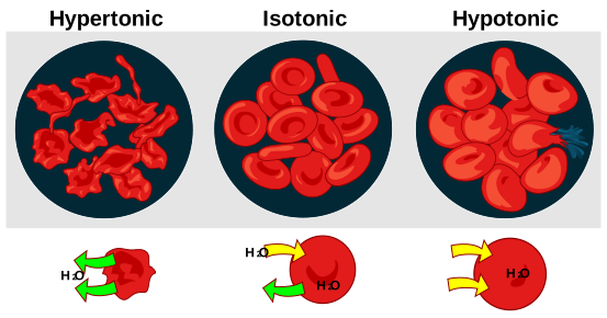
Hypotonic means fewer dissolved particles outside than in. Water rushes into the cell, and since a red blood cell has no wall to stop it, the cell swells until it bursts. Bursting a red blood cell is called haemolysis, and the released haemoglobin then has to be cleared by the kidneys, which they do badly.
Isotonic means the particle concentration matches, so there is no net movement and the cell keeps its shape.
Hypertonic means more particles outside. Water leaves, and the cell shrivels into a spiky shape called a crenated cell.
This is why an intravenous drip is not pure water. Plasma has an osmolality of about 275 to 295 milliosmoles per kilogram. Giving a litre of pure water directly into a vein would be violently hypotonic and would burst red blood cells throughout the circulation. So the standard fluid is normal saline: 0.9 percent sodium chloride, which is 154 millimoles of sodium and 154 of chloride per litre, giving about 308 milliosmoles per litre — close enough to plasma to be safe. Where sugar is preferred, 5 percent dextrose is used, which is isotonic in the bag and then becomes effectively free water as cells consume the glucose.
And it is why drinking sea water kills you faster than drinking nothing. Sea water is around three and a half times more concentrated than your plasma. Your kidney cannot produce urine more concentrated than roughly 1,200 milliosmoles per kilogram, so excreting the salt from a litre of sea water requires more than a litre of water, taken from your own tissues. Every mouthful dehydrates you further. Chapter 23.10 covers survival at sea, where this is the first rule.
Water gets its own doors. Water can trickle through the lipid, but tissues that move serious volumes need more. Aquaporins are channel proteins that pass water and nothing else — a pore narrow enough that even a hydrogen ion cannot get through, with a twist in the middle that flips each water molecule as it passes and breaks any chain that could carry a proton. Peter Agre identified them and shared the 2003 Nobel Prize in Chemistry. Your kidney's ability to concentrate urine depends entirely on inserting and removing aquaporins in the collecting duct on hormonal command, which is Chapter 10.3.
Facilitated diffusion: a door, still downhill
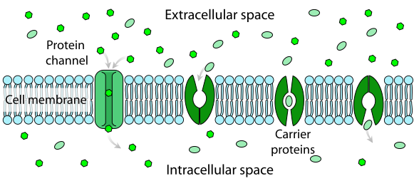
Facilitated diffusion is movement through a protein, down the gradient, at no energy cost. Two kinds of protein do it, both shown in the earlier figure.
Channels are pores. They are fast — millions of ions a second — and usually selective and gated, meaning they open and close in response to a voltage change, a bound molecule, or physical stretch. Every nerve impulse in your body is channels opening and closing in sequence, which is Chapter 11.1.
Carriers bind their passenger, change shape, and release it on the other side. They are slower and can be saturated exactly like an enzyme. GLUT transporters carry glucose into cells this way, and which GLUT a tissue uses is clinically important: GLUT4, in muscle and fat, only appears in the membrane when insulin tells it to, which is the central fact of Chapter 18.7 on diabetes. GLUT2 in the liver and pancreas works without insulin, which is how those organs sense blood sugar directly.
Active transport: paying to go uphill
Everything so far runs downhill. To build a gradient rather than spend one, a cell has to pay, and it pays in ATP.

The sodium–potassium pump is in the membrane of every animal cell you have, and it runs continuously from before you were born until you die. Follow the cycle in the figure.
Three sodium ions from inside the cell bind to the pump. ATP is split, and the phosphate that comes off attaches to the pump itself. That attachment forces the protein into a different shape, one whose sodium sites face outward and grip weakly, so the three sodium ions are released outside. In that new shape, two potassium sites face outward and grip strongly, so two potassium ions from outside bind. Their binding triggers the phosphate to fall off, the pump snaps back to its original shape, and the two potassium ions are released inside.
Three sodium out, two potassium in, one ATP spent. Note that the counts do not match — three positive charges leave and only two come in, so every cycle makes the inside of the cell slightly more negative. A pump that moves net charge is called electrogenic.
This one protein consumes somewhere around 20 to 30 percent of your entire resting energy expenditure. In neurons, which have to reset their ion gradients after every impulse, the figure is far higher — up to about 70 percent of that cell's energy budget. A significant fraction of everything you eat is spent moving sodium out of your cells and potassium in.
And it buys three things.
A membrane potential. The steady leak of potassium outward through always-open channels, combined with the pump's charge imbalance, leaves the inside of a resting cell at about −70 millivolts relative to outside. That stored voltage is what a nerve impulse spends. Chapter 11.1 derives the number.
Cell volume control. A cell is full of proteins and other large molecules that cannot leave, and those molecules pull water in osmotically. Left alone, every cell in your body would swell and burst. Pumping sodium out continuously offsets that pull. This is why cells swell when they run out of oxygen — no ATP, no pump, no defence.
A sodium gradient the cell can spend on something else. This last one is the payoff.
Secondary active transport, and why oral rehydration works
The pump has stored energy in the form of a steep sodium gradient: sodium is desperate to get back in. A second protein can open a door for sodium and charge it a toll — carrying something else uphill on the same trip. That is secondary active transport, and it costs no ATP directly, only the gradient the pump already paid for.
The best example is in the lining of your small intestine. A carrier called SGLT1 binds two sodium ions and one glucose molecule together, and lets the sodium rush in. The energy released drags the glucose in with it, even when there is already more glucose inside the cell than outside. Glucose absorption from your food depends on this. And where glucose and sodium go, water follows by osmosis.
Now put that together with cholera. The cholera bacterium does not invade anything. It releases a toxin that locks open a chloride channel in the gut lining so that chloride, and then sodium, and then water, pour out into the bowel continuously. An adult can lose more than ten litres a day. Untreated, severe cholera kills by dehydration and circulatory collapse in hours, and the historical death rate was around 50 percent.
But the toxin does not touch SGLT1. The absorption route is still working — it just needs sodium and glucose delivered together to run. Give plain water and very little is absorbed. Give water with sodium and glucose in the right ratio and SGLT1 pulls the sodium in, glucose comes with it, and water follows osmotically, faster than the toxin can push it out.
That is oral rehydration solution. The World Health Organization's reduced-osmolarity formula is 75 millimoles per litre of sodium, 75 of glucose, plus potassium and citrate, totalling about 245 milliosmoles per litre. It costs a few cents, needs no needle, no refrigeration and no doctor, and it dropped the death rate of severe cholera from around 50 percent to under 1 percent. The Lancet called it potentially the most important medical advance of the twentieth century. It is a bench-top fact about a carrier protein, turned into a sachet.
Chapter 23.6 gives the home recipe and, just as importantly, the ratio you must not get wrong — too much salt makes the solution hypertonic and pulls water into the bowel, worsening the diarrhoea it was meant to treat.
Two more places the pump appears
Digoxin, a heart drug derived from the foxglove plant, works by partially blocking the sodium–potassium pump in heart muscle cells. Less sodium is pumped out, so the sodium gradient weakens, so a different exchanger that normally uses that gradient to expel calcium works less well, so calcium builds up inside the cell — and calcium is what makes heart muscle contract. The result is a stronger heartbeat. It also explains why digoxin is dangerous: the effective dose and the toxic dose are uncomfortably close, and low blood potassium makes toxicity worse because potassium and digoxin compete for the same site on the pump. Chapter 22.7.
SGLT2 inhibitors — the diabetes drugs whose names end in -flozin — block a relative of SGLT1 in the kidney, the one that normally recovers glucose from urine before it is lost. Block it and glucose leaves in the urine, lowering blood sugar by a route that has nothing to do with insulin. Chapter 22.8.
Bulk transport: when the cargo is too big for any door
Some things are simply too large to go through a protein, so the membrane engulfs them instead.
Endocytosis is the membrane folding inward around something and pinching off an internal bubble. Phagocytosis — cell eating — is the version for large particles, and it is how your white blood cells swallow bacteria whole (Chapter 13.1). Pinocytosis — cell drinking — is the same thing for droplets of fluid.
Receptor-mediated endocytosis is the precise version: receptors on the surface bind one specific molecule, gather together in a coated pit, and the pit pinches off carrying a concentrated load. Your cells take up cholesterol this way, by binding LDL particles with the LDL receptor. Michael Brown and Joseph Goldstein worked this out and received the 1985 Nobel Prize in Medicine, and the reason it mattered is familial hypercholesterolaemia: people with a faulty LDL receptor cannot clear LDL from their blood, run cholesterol levels several times normal from childhood, and have heart attacks decades early. That discovery is also where statins came from.
Exocytosis is the reverse — an internal vesicle fuses with the membrane and dumps its contents outside. This is how insulin leaves a pancreatic beta cell, how a neuron releases neurotransmitter into a synapse, and how digestive enzymes leave the pancreas.
When a channel is the disease
Cystic fibrosis is caused by a faulty chloride channel called CFTR. Chapter 1.3 covered the folding failure; here is the consequence. Without working CFTR, cells lining the airways cannot move chloride out, so water does not follow, so the mucus layer on the airway surface becomes thick and sticky instead of thin and mobile. The tiny hairs that normally sweep mucus upward cannot move it. Bacteria settle in and are never cleared, and repeated infection destroys the lungs. The same failure blocks the pancreatic ducts, which is why these patients cannot digest fat properly and need enzyme capsules with every meal.
It also produces the oldest diagnostic test in the disease and one of the most curious. CFTR in sweat glands normally recovers chloride from sweat before it reaches the skin. Without it, the chloride stays, and the sweat is salty enough to taste. A northern European folk saying, recorded in the 1600s, held that a child whose forehead tastes of salt when kissed is bewitched and will soon die. The sweat chloride test is still the standard diagnostic method, three and a half centuries later.
What the next page fixes
This page treated the cell as one compartment with a boundary. It is not. Inside that boundary are a dozen further compartments, each sealed off by its own membrane and running its own chemistry — a nucleus, a set of power stations, a production line, a recycling plant and a waste disposal system. Chapter 1.5 opens the cell and goes through them one at a time, including what disease each one causes when it fails.