Appearance
12.2 — The Hypothalamus and Pituitary
The pituitary gland weighs about half a gram and is the size of a pea. It sits in a bony hollow at the base of the skull, hanging from the brain on a stalk, and it controls the thyroid, the adrenals, growth, reproduction, lactation, and water balance.
It was called the master gland for a century. That title now belongs to the hypothalamus above it, which controls the pituitary — and which is itself under the influence of the rest of the brain, so the chain of command runs back into thought and emotion.
Two glands in one
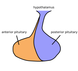
The anterior pituitary is glandular tissue, derived embryologically from the roof of the mouth, which grew upward to meet the developing brain.
It is controlled by hormones, delivered through a small private circulation — the hypophyseal portal system — running down the stalk. A portal system is one that runs capillary bed to capillary bed without returning to the heart in between (Chapter 7.5).
And that arrangement matters enormously. It means hypothalamic hormones reach the pituitary at high concentration, in tiny total quantities, without being diluted in the whole circulation.
The posterior pituitary is not a gland at all. It is nervous tissue — the axon terminals of neurons whose cell bodies are in the hypothalamus.
It manufactures nothing. The hormones are made in the hypothalamus, transported down the axons, stored in the terminals, and released into the blood on a nerve impulse.
So it is a place where nerve endings secrete into the bloodstream instead of onto another cell — which is exactly what the adrenal medulla does (Chapter 4.4), and it is the clearest illustration that the nervous and endocrine systems are not really separate.
And this explains a clinical pattern. Damage to the stalk cuts off the anterior lobe's blood-borne control and cuts the posterior lobe's axons. So a stalk lesion causes deficiency of anterior hormones and diabetes insipidus together.
The hypothalamus
About 4 grams, sitting below the thalamus and above the pituitary, and controlling an implausible range of functions (Chapter 11.4).
Its releasing and inhibiting hormones, each controlling one pituitary hormone:
| Hypothalamic hormone | Effect on pituitary |
|---|---|
| TRH | Stimulates TSH and prolactin |
| CRH | Stimulates ACTH |
| GnRH | Stimulates LH and FSH |
| GHRH | Stimulates growth hormone |
| Somatostatin | Inhibits growth hormone and TSH |
| Dopamine | Inhibits prolactin |
That last row is the odd one out and it has real consequences.
Prolactin is the only pituitary hormone under predominantly inhibitory control. Everything else needs a signal to be released; prolactin is continuously suppressed by dopamine and is released when the suppression stops.
So anything that cuts the stalk, or blocks dopamine, raises prolactin.
This is why antipsychotics cause milk production and breast enlargement — they block dopamine receptors, including the ones suppressing prolactin (Chapter 11.2). And it is why a stalk lesion causes every pituitary hormone to fall except prolactin, which rises. A mildly raised prolactin with low everything else points to a stalk problem rather than a prolactin-secreting tumour.
And it works in reverse therapeutically: dopamine agonists such as cabergoline reduce prolactin, and they are the first-line treatment for a prolactin-secreting tumour — a case where a tumour is treated with tablets rather than surgery, and shrinks.
The anterior pituitary hormones
Six, and a memorable grouping: four are tropic (controlling other glands) and two act directly.
TSH — thyroid-stimulating hormone. Controls the thyroid (Chapter 12.3).
ACTH — adrenocorticotropic hormone. Controls cortisol production by the adrenal cortex (Chapter 12.4).
And ACTH has a side effect worth knowing. It is made from a larger precursor protein that also yields melanocyte-stimulating hormone, and ACTH itself weakly stimulates pigment cells. So very high ACTH causes skin darkening, particularly in skin creases, scars and the inside of the mouth. This is the tanned appearance of Addison's disease — a person who appears to have been on holiday and is in fact adrenally insufficient.
LH and FSH — the gonadotropins. Control the ovaries and testes (Chapters 15.2, 15.4).
Growth hormone. Acts directly on tissues, and indirectly through IGF-1 made by the liver.
Prolactin. Milk production.
Growth hormone deserves more detail because its excess and deficiency are both distinctive.
It promotes growth of bone and soft tissue, increases protein synthesis, mobilises fat, and raises blood glucose — it is a counter-regulatory hormone, opposing insulin.
Deficiency in a child causes short stature with normal proportions. Treated with growth hormone injections, and effective if started early.
Excess in a child, before the growth plates close (Chapter 5.1), causes gigantism.
Excess in an adult, after the plates have closed, causes acromegaly — and the difference is entirely about whether the plates are open.
Acromegaly is one of the diseases most reliably missed for years, because it develops so gradually that neither the patient nor their family notices.
Bones cannot lengthen, so they thicken. The hands and feet enlarge — people notice that rings no longer fit and shoe size has increased in adulthood. The jaw protrudes, the brow ridge thickens, the nose and lips enlarge, and the teeth become spaced apart.
And the diagnosis is often made from old photographs, comparing the face over a decade. A doctor asking to see photographs from ten years ago is doing exactly the right thing.
The systemic effects are what shorten life: hypertension, cardiomyopathy, diabetes, sleep apnoea, arthritis and increased bowel cancer risk. Treated, life expectancy is close to normal; untreated, it is reduced by about ten years.
Diagnosis uses the suppression principle (Chapter 12.1). Growth hormone should be suppressed by glucose; in acromegaly it is not. Treatment is surgery, usually through the nose (below), with somatostatin analogues and radiotherapy as further options.
The posterior pituitary hormones
Two, both made in the hypothalamus.
ADH (vasopressin) — water retention (Chapter 10.2), and vasoconstriction at high concentrations.
Oxytocin — uterine contraction in labour and milk ejection in breastfeeding (Chapter 15.7).
The two are structurally almost identical — nine amino acids each, differing in two. Which is why oxytocin has some antidiuretic activity, and why high-dose oxytocin infusions in labour can cause water retention and dangerous hyponatraemia if given in large volumes of dilute fluid.
Oxytocin's popular reputation as "the love hormone" runs far ahead of the evidence. It has clear, well-established roles in labour, lactation and maternal behaviour. Studies of intranasal oxytocin and human trust or social behaviour have produced inconsistent results and have replicated poorly, and the confident claims made about it in popular writing should be treated with scepticism.
Pituitary disease
The gland's position explains almost everything about how it presents.
It sits in a bony hollow — the sella turcica — with the optic chiasm immediately above it (Chapter 11.7).
So a pituitary tumour has two ways of causing symptoms: by producing hormone, and by pressing on things.
And the pressure symptom is the classic one. The chiasm carries the crossing fibres from the nasal half of each retina, which serve the outer half of each visual field. A tumour growing upward compresses them first.
The result is bitemporal hemianopia — loss of the outer half of the visual field in both eyes.
And it is frequently unnoticed for a long time, because the person still has central vision and the brain fills in. They bump into things on both sides, or have near-misses when driving, and attribute it to carelessness. Formal field testing reveals it immediately.
Most pituitary tumours are benign adenomas, and they are classified by what they secrete.
Prolactinoma — the commonest. In women it causes absent periods, milk production and infertility, so it is usually diagnosed while small. In men it causes reduced libido and erectile dysfunction, which are attributed to other things, so it is usually large by the time it is found — a striking example of how presentation shapes diagnosis. Treated with dopamine agonists, which shrink it.
Growth hormone-secreting — acromegaly, above.
ACTH-secreting — Cushing's disease (Chapter 12.4).
Non-functioning — presenting purely by pressure.
Hypopituitarism — deficiency of several hormones. The classic order of loss as a tumour grows is growth hormone first, then the gonadotropins, then TSH, then ACTH. So reduced libido and fatigue precede the more dramatic features.
Sheehan's syndrome is a specific and tragic cause. The pituitary enlarges substantially in pregnancy, increasing its blood demand; severe postpartum haemorrhage can then infarct it. The first sign is usually failure to lactate, followed by failure of periods to return, and then the features of thyroid and adrenal deficiency. It is now rare where obstetric haemorrhage is well managed, and it remains a real cause where it is not.
Pituitary apoplexy — sudden haemorrhage or infarction into a pituitary tumour. Sudden severe headache, visual loss, eye movement problems, and collapse from acute adrenal insufficiency. It is an emergency, and the immediate treatment is steroid replacement, before any imaging or surgery, because the cortisol deficiency is what kills.
Empty sella — the hollow appears empty on a scan because the gland is flattened. Usually an incidental finding in someone with normal pituitary function, and it causes unnecessary alarm when reported without explanation.
Trans-sphenoidal surgery
Most pituitary surgery is done through the nose, passing through the sphenoid sinus (Chapter 5.2) into the sella from below.
No incision, no brain retraction, and a hospital stay of a few days. It is a genuinely elegant piece of anatomical opportunism: the sinus happens to sit directly beneath the gland, and it is already an air-filled space open to the outside.
Increasingly it is done endoscopically, with a camera passed through the nostril.
The complications follow the anatomy. Damage to the posterior pituitary or stalk causes diabetes insipidus, often transient. A cerebrospinal fluid leak can occur if the covering above the gland is breached, and it presents as clear fluid dripping from the nose. And the carotid arteries run in the walls of the cavity on either side.
Diabetes insipidus
Covered in Chapter 10.2, and the key clinical points belong here.
Enormous volumes of dilute urine — up to 20 litres a day — with constant thirst.
Distinguishing it from psychogenic polydipsia — compulsive water drinking — is done with a water deprivation test. Deprive of water: a person with diabetes insipidus continues producing dilute urine and becomes dehydrated; a compulsive drinker concentrates their urine normally.
Then give desmopressin. The central form responds; the nephrogenic form does not.
Treatment of the central form is desmopressin, a modified ADH given as a nasal spray or tablet.
And its most common everyday use is quite different: desmopressin is used for bedwetting in children, reducing overnight urine production. It is effective and it must be used with fluid restriction in the evening, because taking it while drinking freely can cause dangerous hyponatraemia — a genuine and occasionally fatal complication.
What the next page fixes
The pituitary's largest single target sets the metabolic rate of every cell you have. Chapter 12.3 covers the thyroid and parathyroids — one gland controlling how fast you run and, wrapped around it, four the size of grains of rice controlling your blood calcium.