Appearance
6.3 — Muscles of the Head and Neck
Almost every skeletal muscle in your body runs from one bone to another. The muscles of facial expression are the exception: they attach to skin. That is why they move the face rather than the skeleton, and it is why a single nerve injury produces one of the most instantly recognisable clinical pictures in medicine.
The head and neck contain three functional groups: the muscles of expression, the muscles of chewing, and the muscles of the neck that move the head and control swallowing.
Muscles of facial expression
There are more than twenty on each side. All of them are supplied by the facial nerve — cranial nerve VII — and all of them derive from the same embryonic structure, which is why one nerve controls the whole set.
Orbicularis oculi — a ring around the eye. Closes the eyelids, gently for blinking and forcefully for screwing the eyes shut. The blink reflex protects the cornea, and it matters more than it sounds: the cornea has no blood supply and is kept moist and clear entirely by tear film spread by blinking.
Orbicularis oris — a ring around the mouth. Closes and purses the lips. Essential for speech, whistling, drinking through a straw, and keeping food in the mouth.
Buccinator — in the cheek. Presses the cheek against the teeth, which keeps food between the grinding surfaces rather than collecting in the space between cheek and gums. It also generates the pressure for blowing, and its name comes from the Latin for trumpeter.
Zygomaticus major — from the cheekbone to the corner of the mouth. Pulls the corner up and back. This is the smile muscle.
Frontalis — over the forehead. Raises the eyebrows and wrinkles the forehead.
Corrugator supercilii — draws the eyebrows together, producing the vertical frown lines. This is the muscle most commonly injected with botulinum toxin for cosmetic purposes (Chapter 6.2).
Platysma — a broad thin sheet in the neck, continuous with the facial muscles. Tenses the skin of the neck.
And there is a distinction worth knowing about smiles. A genuine smile involves both zygomaticus major and orbicularis oculi, so the eyes crease. A voluntary social smile usually involves only the mouth, because orbicularis oculi is much harder to contract deliberately. The observation is credited to Guillaume Duchenne in the nineteenth century, and the "Duchenne smile" is still the term used. It is not infallible — some people can produce it voluntarily — but it is a real and measurable difference.
Facial nerve palsy
Damage to the facial nerve paralyses one entire side of the face, and the picture is unmistakable: the forehead does not wrinkle, the eye cannot close, the nasolabial fold flattens, the mouth droops, and speech and drinking are affected on that side.
Bell's palsy is the commonest form — sudden, one-sided, and idiopathic, though reactivation of herpes simplex virus in the nerve's ganglion is the leading explanation. It affects around 1 in 60 people at some point in life. Around 70 percent recover completely without treatment, and 85 percent with treatment; steroids started within 72 hours improve the odds, and the earlier the better.
The single most important immediate concern is the eye. The person cannot blink and cannot close the eye, so the cornea dries out and can ulcerate and perforate. Artificial tears during the day, ointment and taping the eye shut at night — this is what protects the sight, and it is frequently the part that gets least attention.
And there is one distinction that must be made in every case, because it separates a benign condition from a stroke.
The forehead has bilateral cortical supply. The part of the brain controlling the lower face sends fibres only to the opposite side; the part controlling the forehead sends fibres to both sides.
So:
A stroke affecting the face spares the forehead. The person cannot smile on one side, but they can still raise both eyebrows and wrinkle their forehead, because the surviving hemisphere still supplies it.
A facial nerve palsy affects the forehead too, because the damage is beyond where the two supplies have merged.
"Can you raise your eyebrows?" therefore distinguishes a Bell's palsy from a stroke in about three seconds, and it is one of the highest-value questions in all of clinical examination. A drooping face with a working forehead is a stroke until proven otherwise, and that is a time-critical emergency (Chapter 18.6).
Ramsay Hunt syndrome is facial palsy caused by shingles in the facial nerve's ganglion. Look in the ear: a vesicular rash in the ear canal or on the eardrum is the giveaway, and it needs antivirals as well as steroids. It has a worse prognosis than Bell's palsy, and missing it costs recovery.
Muscles of mastication
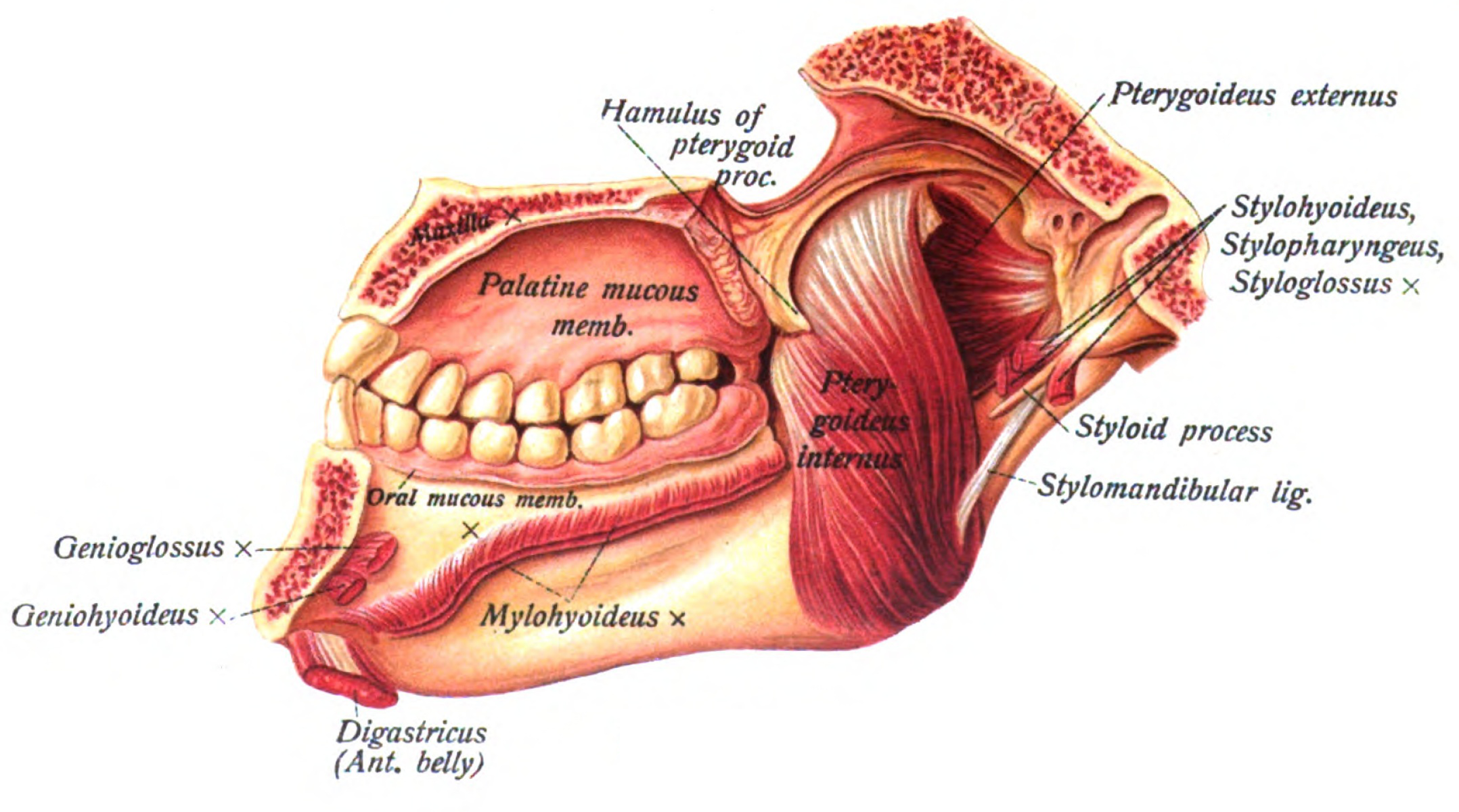
Four on each side, all supplied by the trigeminal nerve — cranial nerve V — which is a different nerve entirely from the one supplying expression. This separation is why a facial nerve palsy does not affect chewing.
Masseter — from the cheekbone to the angle of the jaw. The strongest muscle in the body by weight, and you can feel it bulge by clenching your teeth. Bite force at the molars reaches around 700 newtons in an adult.
Temporalis — a broad fan filling the hollow at the temple, running down under the cheekbone arch to the jaw. Closes the jaw and retracts it.
Medial and lateral pterygoid — deep muscles between the jaw and the skull base. The medial helps close; the lateral is the one that opens the jaw and moves it side to side for grinding.
The temporomandibular joint is unusual: it both hinges and slides. Opening the mouth involves the jaw rotating and then translating forward. This is why the jaw can dislocate on a wide yawn — the condyle slides forward past the point it should stop and locks there, and reduction requires pushing down and back.
Temporomandibular joint dysfunction — pain around the joint, clicking, restricted opening, and headache — is common, frequently related to nocturnal grinding, and mostly managed conservatively with a bite splint, jaw rest and stress management rather than surgery.
And jaw claudication is a red flag. Pain in the jaw that comes on while chewing and eases with rest, in someone over 50, is the classic symptom of giant cell arteritis, in which inflamed arteries cannot supply enough blood to the working muscle. It matters because the same disease can occlude the artery supplying the eye and cause sudden irreversible blindness, and it is treated with high-dose steroids the same day, before biopsy confirmation. Chapter 21.5.
The neck

Sternocleidomastoid — the name is a list of its attachments: sternum, clavicle, mastoid process. It runs diagonally across the side of the neck.
Its action is worth working out rather than memorising. Contracting one side turns the head to the opposite side and tilts it toward the same side. Contracting both together flexes the neck — and, with the head fixed, helps lift the ribcage in laboured breathing.
That last role is a clinical sign. A person using their sternocleidomastoids visibly to breathe is in respiratory distress, and the use of accessory muscles is one of the fastest ways to judge severity of an asthma attack or heart failure from across a room (Chapter 8.3).
Congenital torticollis is a shortened sternocleidomastoid in an infant, from positioning in the womb or birth injury, producing a head tilted to one side. Physiotherapy and stretching in the first months resolves most cases, and delay makes it harder to correct.
Scalene muscles — three on each side, running from the cervical vertebrae to the first two ribs. They elevate the ribs in inspiration and bend the neck sideways. The brachial plexus and the subclavian artery pass between two of them, and compression at that point causes thoracic outlet syndrome, with arm pain, numbness and sometimes vascular symptoms.
The strap muscles — thin muscles in front of the trachea that raise and lower the larynx during swallowing and speech.
Trapezius — a large diamond-shaped muscle spanning the back of the neck and upper back, attaching to the skull, the spine and the shoulder girdle. It shrugs the shoulders and rotates the scapula upward. It is supplied by the accessory nerve — cranial nerve XI — which runs superficially through the neck, and this is a genuine surgical hazard: the nerve is easily damaged during lymph node biopsy in the neck, and the result is a shoulder that cannot be lifted properly and chronic pain.
Swallowing
Swallowing involves more than 30 muscles, coordinated by a centre in the brainstem, and it happens 500 to 700 times a day, mostly without any awareness.
Three phases:
Oral (voluntary) — the tongue pushes the food bolus back against the palate.
Pharyngeal (reflex, under a second) — and this is where the danger sits. The soft palate rises to seal the nose. The larynx lifts and moves forward, and the epiglottis folds back over the airway. The vocal cords close. Breathing stops. Then the muscles of the pharynx contract in sequence, pushing the bolus down, and the upper oesophageal sphincter relaxes to let it through.
Oesophageal — a wave of muscular contraction carries the bolus to the stomach in about 8 to 10 seconds.
Every element of the pharyngeal phase exists to protect the airway, because of the crossing of air and food passages described in Chapter 3.6. It is a reflex with no margin for error, run several hundred times a day.
Dysphagia and aspiration
When swallowing fails, food and liquid enter the airway — aspiration. The consequences are large and frequently underestimated.
Stroke is the commonest cause, affecting around half of stroke patients acutely. Aspiration pneumonia is a leading cause of death after stroke, and it kills people who survived the stroke itself.
This is why every stroke patient is given a swallow assessment before anything is given by mouth, and why "nil by mouth until swallow screened" is one of the standard immediate orders. It is not caution for its own sake — it is a protocol that measurably reduces mortality.
Silent aspiration is the dangerous version: material enters the airway without producing a cough, because the sensation that triggers the cough reflex has also been lost. The absence of coughing is not evidence of safe swallowing, which is exactly why formal assessment is used rather than watching someone drink a glass of water.
Other causes: Parkinson's disease, motor neurone disease, myasthenia gravis, oesophageal cancer or stricture, and simply the reduced reflex efficiency of advanced age.
And the pattern of the difficulty localises the problem. Difficulty with liquids more than solids suggests a neurological or muscular problem, because thin liquids move fast and need precise timing. Progressive difficulty with solids first, then liquids, suggests a narrowing — a stricture or a tumour — and that pattern in an adult, particularly with weight loss, warrants urgent endoscopy (Chapter 21.2).
What the next page fixes
The head sits on a trunk whose muscles do something quite different from moving bones: they contain the abdominal contents, generate pressure for coughing, childbirth and defecation, and stabilise the spine before any limb moves. Chapter 6.4 covers the trunk and core, including what the diaphragm actually does and why "core stability" is more specific and more useful than the fitness-industry version of it.