Appearance
5.2 — The Skull
The skull is 22 bones — 8 forming the braincase and 14 forming the face — plus the six tiny bones of the middle ears and the hyoid in the neck, which touches no other bone at all.
It does three incompatible jobs at once. It has to be a rigid armoured box around a brain that cannot be compressed. It has to be full of holes for the eyes, nose, ears, mouth and every nerve and vessel entering or leaving the head. And it has to be light, because it sits on top of a vertical spine and every gram costs balance and muscle work.
The compromises it makes are why head injuries behave the way they do.
The braincase

Frontal — the forehead and the roofs of the eye sockets. Contains the frontal sinuses.
Parietal (two) — the sides and roof.
Temporal (two) — the sides around the ears. This bone contains the entire hearing and balance apparatus sealed inside its densest portion, the petrous part, which is the hardest bone in the body and frequently the last part of a skeleton to decay.
Occipital — the back and base. It carries the foramen magnum, the large hole through which the brainstem becomes the spinal cord, and the two rounded surfaces on which the skull pivots on the first vertebra.
Sphenoid — a single bone shaped like a bat with outstretched wings, wedged across the base of the skull and articulating with almost every other cranial bone. It carries a saddle-shaped hollow, the sella turcica, in which the pituitary gland sits (Chapter 12.2).
Ethmoid — a delicate bone between the eye sockets, forming part of the nasal cavity roof. Its cribriform plate is a thin sheet perforated by the fibres of the olfactory nerve.
That plate is one of the most dangerous structures in the skull. It is paper-thin, and a blow to the front of the head can fracture it. When it breaks, three things follow: the olfactory fibres are torn, so smell is lost, often permanently; cerebrospinal fluid can leak from the nose, appearing as clear fluid dripping from one nostril; and there is now a direct route from the nasal cavity to the space around the brain, which is a path for meningitis. Clear fluid running from the nose after a head injury is a serious sign, not a runny nose, and it is why patients with head injuries are asked about it directly.
The sutures
The cranial bones meet at sutures — interlocking joints with a thin layer of fibrous tissue between them, immovable in an adult.
In an infant they are not fused, and that is deliberate. Unfused plates let the head compress and overlap during birth, which is what makes passage through the pelvis possible (Chapter 3.6), and they allow the skull to grow with the brain, which triples in volume in the first year.
The gaps between them are the fontanelles. The anterior fontanelle, at the top front, is the large diamond-shaped soft spot; it closes between 9 and 18 months. The posterior one closes by about 2 months.
The anterior fontanelle is a clinical window and it is genuinely useful. In a calm infant held upright it should be flat and soft. A bulging, tense fontanelle means raised intracranial pressure — meningitis, bleeding, hydrocephalus — and it is one of the most valuable signs in paediatrics because it can be checked in seconds without any equipment. A sunken fontanelle means dehydration.
Premature fusion of a suture is called craniosynostosis. The skull cannot grow across that suture, so it grows excessively in the perpendicular direction, and the head takes a characteristic shape depending on which suture closed. Surgery is needed both for appearance and, when several sutures fuse, to prevent the brain being constrained.
The face
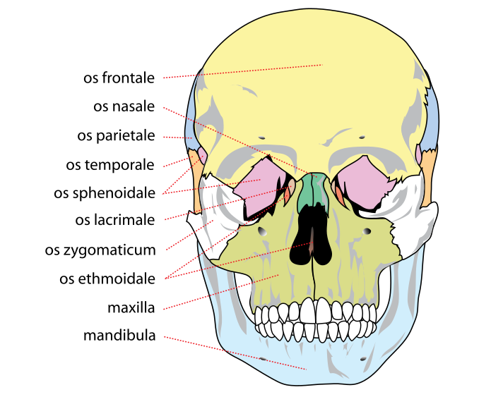
Maxilla (two, fused) — the upper jaw, holding the upper teeth, forming the floor of each eye socket, the sides of the nose, and most of the hard palate. Contains the maxillary sinuses.
Zygomatic (two) — the cheekbones, forming a prominent arch that is easily felt and easily broken.
Nasal (two) — the bridge of the nose. The commonest fractured bone in the face, and the reason is that it projects further than anything else.
Mandible — the lower jaw, and the only skull bone that moves. Its joint with the temporal bone is the temporomandibular joint.
Plus the lacrimal bones (tear ducts), palatine bones (back of the hard palate), inferior nasal conchae, and the vomer, which forms the lower part of the nasal septum.
The hyoid sits in the front of the neck, suspended by muscles and ligaments, touching no other bone. It anchors the tongue and the muscles of swallowing. A fractured hyoid at post-mortem is strongly suggestive of manual strangulation, because very few other mechanisms apply enough force to it, and it is one of the classic findings in forensic pathology.
The sinuses
Four paired air-filled cavities within skull bones, all connected to the nasal cavity: frontal, maxillary, ethmoid and sphenoid.
What they are for is genuinely uncertain. The commonly repeated explanations — lightening the skull, warming inhaled air, adding resonance to the voice — are all plausible and none is well established. The honest answer is that nobody knows, which is worth saying rather than repeating a confident guess.
What is certain is the trouble they cause. Each drains through a narrow opening, and if that opening blocks — from a cold, an allergy, or a deviated septum — the sinus fills with fluid, bacteria multiply, and you have sinusitis: facial pain worsened by leaning forward, blocked nose, and a discharge.
And the maxillary sinus is the worst-designed of them, for the reason given in Chapter 3.6: its drainage opening is near its roof. In a quadruped with a horizontal head that is the low point. Standing upright made it the high point, so fluid must be moved upward against gravity by cilia alone. This is a direct legacy of walking upright, and it is why maxillary sinusitis is the commonest and most stubborn.
The dangerous part is what the sinuses are next to. The ethmoid sinuses are separated from the eye socket by a wall of bone under a millimetre thick, so ethmoid infection can spread into the orbit — orbital cellulitis, which threatens vision and can progress to the brain, and which is an emergency requiring intravenous antibiotics. The frontal and sphenoid sinuses sit directly against the front of the brain and the pituitary respectively.
And that proximity is also exploited surgically. A pituitary tumour is usually removed by going through the nose and the sphenoid sinus — the trans-sphenoidal approach — reaching the gland without opening the skull at all. Chapter 12.2.
The base of the skull, and what passes through it
The floor of the cranial cavity is stepped into three levels and is perforated by openings carrying every nerve and vessel between the brain and the rest of you.
The foramen magnum carries the brainstem, the vertebral arteries and the spinal accessory nerves.
The optic canal carries the optic nerve and ophthalmic artery.
The jugular foramen carries the internal jugular vein and three cranial nerves.
And the carotid canal carries the internal carotid artery, which enters the skull through the temporal bone.
Base of skull fractures produce signs that are worth knowing because they are visible from across a room.
Battle's sign — bruising over the mastoid process, behind the ear, appearing 12 to 24 hours after injury. Raccoon eyes (periorbital ecchymosis) — bruising around both eyes with no direct injury to them. Cerebrospinal fluid leaking from the nose or the ear.Blood behind the eardrum.
Any of these means a base of skull fracture, and the delay before the bruising appears is why a patient who looked fine at the scene can look alarming the next morning.
The two head injuries you should be able to recognise
Extradural (epidural) haematoma. The middle meningeal artery runs in a groove on the inside of the temporal bone, and the temporal bone is the thinnest part of the skull vault. A blow to the side of the head fractures it and tears the artery. Arterial blood then strips the tough outer membrane away from the inside of the skull and accumulates under pressure.
The classic pattern is the reason this is famous: a blow, brief loss of consciousness, then a lucid interval of minutes to hours in which the person seems recovered and may even walk and talk, followed by rapid deterioration into coma. The lucid interval happens because it takes time for enough blood to accumulate to compress the brain.
It is a surgical emergency with an excellent outcome if operated on early and a very poor one if not. This is precisely why head injury advice sheets tell you to return if symptoms develop hours later, and why someone with a significant head injury should not be left alone to sleep unobserved.
Subdural haematoma is different in almost every respect. It comes from tearing the small bridging veins that cross from the brain surface to the venous sinuses. Venous bleeding is slow, so it can accumulate over days to weeks. It is commonest in the elderly and in people with alcohol dependence, because a brain that has shrunk slightly puts more stretch on those veins, and it can follow an injury so minor the person does not recall it. The presentation is often gradual confusion, headache or personality change rather than dramatic collapse, which is exactly why it gets missed and put down to dementia.
The teeth
Adults have 32 permanent teeth — per quadrant: two incisors for cutting, one canine for tearing, two premolars and three molars for grinding. Children have 20 deciduous teeth.
Enamel is the hardest substance in the body, about 96 percent mineral. It contains no cells and no blood supply, so it cannot repair itself. A cavity does not heal. This is the single most important fact about dental disease and the entire reason prevention matters more here than almost anywhere else in medicine.
Underneath is dentine, softer, containing fine tubules that run to the pulp. Once decay reaches dentine, the tubules transmit stimuli to the nerve, which is why a cavity becomes sensitive to cold and sweet things.
The pulp contains the nerve and blood supply. Infection reaching it causes severe pain and requires either root canal treatment or extraction.
Fluoride works by chemistry. It substitutes into the hydroxyapatite crystal, replacing the hydroxide group and forming fluorapatite, which is measurably more resistant to acid dissolution. Water fluoridation reduces cavities by roughly 25 percent, and it is one of the most cost-effective public health measures available. Excess causes fluorosis — mottling of the enamel — which is why the concentration is regulated rather than maximised.
Wisdom teeth are the third molars, erupting from about 17 to 25. They frequently have nowhere to go, for the reason in Chapter 3.6: the human jaw has shortened faster than the teeth have reduced.
And teeth are the most durable identifier a body has. Enamel survives fire and burial better than bone, dental records are widely kept, and the pattern of fillings and extractions is close to unique. Dental identification is standard in disaster victim identification (Chapter 2.10).
What the next page fixes
The skull sits on a column of 33 vertebrae that carries the entire weight of the upper body, protects the spinal cord, allows movement in every direction, and does all three despite being loaded in a direction it was never built for. Chapter 5.3 covers the spine, the intervertebral disc, and the specific mechanics behind the back pain that four out of five people will experience.