Appearance
11.3 — The Spinal Cord and Reflexes
Touch something hot and your hand is moving before you feel the pain. The withdrawal is complete before the signal has reached your brain at all — the decision was made in your spinal cord, and your brain is informed afterwards.
That arrangement is not a shortcut. It is the reason you still have a hand.
The cord
About 45 centimetres long and 1 to 1.5 centimetres wide, running from the base of the brainstem to about L1 or L2 (Chapter 5.3).
Two enlargements, at the cervical and lumbar levels, where the extra neurons supplying the limbs are located.
31 pairs of spinal nerves: 8 cervical, 12 thoracic, 5 lumbar, 5 sacral, 1 coccygeal.
Note that there are 8 cervical nerves and only 7 cervical vertebrae. C1 to C7 emerge above their corresponding vertebra; C8 emerges below C7; and from T1 down, each nerve emerges below its vertebra. This changes the numbering relationship at that point, and it matters when localising a lesion.
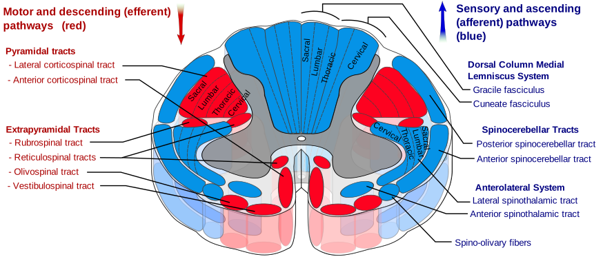
Grey matter in the middle, shaped like a butterfly — cell bodies, dendrites and synapses.
The dorsal (posterior) horn receives sensory input. The ventral (anterior) horn contains motor neurons. The lateral horn, in the thoracic and upper lumbar regions only, contains sympathetic neurons.
White matter around it — myelinated tracts running up and down. White because of the myelin.
Note the inversion compared with the brain, where grey matter is on the outside and white inside.
Each nerve has two roots, and the separation is complete.
The dorsal root carries sensory fibres in, with their cell bodies in a swelling called the dorsal root ganglion, outside the cord. The ventral root carries motor fibres out.
This is the Bell–Magendie law, established in the early nineteenth century, and it is one of the foundational facts of neurology: sensory in at the back, motor out at the front.
And the dorsal root ganglion is where the shingles virus hides. Chickenpox virus travels up a sensory nerve and lies dormant in that ganglion for decades. When it reactivates it travels back down that one nerve, producing pain and a rash confined to exactly one dermatome, stopping abruptly at the midline (Chapter 11.8).
The tracts
Three matter, and knowing them lets you localise a spinal injury from the examination alone.
The dorsal columns — fine touch, vibration, position sense
Enter the cord and ascend on the same side, all the way to the medulla, where they synapse and cross to the other side before continuing to the thalamus and cortex.
So a lesion in the cord affects the same side of the body.
Position sense (proprioception) is the one people underestimate. Close your eyes and you still know where your limbs are — that is this pathway. Lose it and you cannot walk in the dark or with your eyes closed, because vision was compensating.
The test is Romberg's sign: stand with feet together, eyes open, then closed. Marked unsteadiness only with eyes closed indicates loss of position sense, because vision was substituting for it. Unsteadiness with eyes open too points to the cerebellum instead (Chapter 11.4).
Vitamin B12 deficiency damages this pathway specifically — subacute combined degeneration of the cord — which is why B12 deficiency causes numbness, tingling and unsteadiness, and why it must be corrected before folate (Chapter 7.1).
The spinothalamic tract — pain and temperature
Enter the cord, synapse immediately, and cross to the opposite side within one or two segments, then ascend.
So a lesion in the cord affects the opposite side of the body, one or two levels below.
That difference — dorsal columns cross in the medulla, spinothalamic crosses in the cord — is the entire basis of localising spinal lesions, and it produces one of the most striking patterns in clinical medicine.
Brown–Séquard syndrome — damage to one half of the cord. The person loses fine touch and position sense on the same side as the injury, and pain and temperature on the opposite side, both below the level.
It looks bizarre and it is completely predictable once you know where each tract crosses. It is most often seen after a stab wound.
The corticospinal tract — voluntary movement
Descends from the motor cortex, crosses in the medulla, and continues down the cord to synapse on motor neurons.
So the left brain controls the right side of the body. This is why a stroke in one hemisphere weakens the opposite side (Chapter 18.6).
Upper and lower motor neuron lesions produce opposite pictures, and the distinction is one of the most useful in neurology.
| Upper motor neuron | Lower motor neuron | |
|---|---|---|
| Site | Brain or cord | Anterior horn cell, root, or nerve |
| Tone | Increased (spastic) | Decreased (flaccid) |
| Reflexes | Exaggerated | Absent or reduced |
| Plantar response | Upgoing (Babinski) | Downgoing |
| Wasting | Late, from disuse | Early and marked |
| Fasciculations | Absent | Present |
Why an upper motor neuron lesion increases tone is worth explaining, because it is counter-intuitive. The descending pathways are largely inhibitory to the spinal reflex circuits. Remove that inhibition and the reflexes become unrestrained — hence spasticity and brisk reflexes. The cord is not damaged; it has been released from supervision.
The Babinski sign — stroking the sole causes the big toe to go up instead of down. It is normal in infants under about one year, before the corticospinal tract has myelinated, and its return in an adult indicates damage to that tract. A reflex that is normal at six months and pathological at thirty is a direct readout of myelination.
And motor neurone disease is the disease that affects both, producing a mixture of spastic and flaccid features in the same person — which is exactly what makes it recognisable (Chapter 20.6).
Reflexes
A reflex arc has five components: receptor, sensory neuron, integration centre, motor neuron, effector.
Its virtue is speed — no brain involvement, so no delay.
The stretch reflex

The only monosynaptic reflex in the body — one sensory neuron, one motor neuron, one synapse.
Tapping the tendon stretches the muscle. Muscle spindles detect the stretch. The signal enters the cord and synapses directly on the motor neuron for that same muscle, which contracts.
Total time about 20 to 30 milliseconds.
Its real purpose is postural. It resists unexpected lengthening, so when you carry something and it becomes heavier, the muscle stretches slightly and the reflex immediately increases its contraction. You do not decide to grip harder.
And this is why it is tested. The reflex tests a whole segment at once — the sensory nerve, the cord segment, the motor nerve and the muscle.
| Reflex | Root level |
|---|---|
| Biceps | C5, C6 |
| Brachioradialis | C6 |
| Triceps | C7 |
| Knee | L3, L4 |
| Ankle | S1 |
An absent reflex points to a lower motor neuron problem at that level. An exaggerated one points to an upper motor neuron problem above it. Two taps of a hammer, and you have localised a lesion to a segment.
Reinforcement — asking the patient to clench their teeth or pull their interlocked hands apart while you tap — genuinely brings out reflexes that appeared absent, by reducing descending inhibition. An absent reflex is not recorded as absent until reinforcement has been tried.
The withdrawal reflex
Polysynaptic — involving interneurons.
A painful stimulus activates flexors and inhibits extensors on that side, pulling the limb away.
And simultaneously, through interneurons crossing to the other side, it extends the opposite limb — the crossed extensor reflex. If you tread on something sharp, one leg lifts and the other straightens to take your weight. Both happen before you know anything about it.
Other reflexes
The Golgi tendon reflex — tension receptors in the tendon inhibit their own muscle when force becomes very high. This is what stretching exploits to stop a cramp (Chapter 6.7).
Autonomic reflexes — bladder, bowel and sexual reflexes are organised at the sacral level, which is why cord injury above that level leaves the reflexes intact but disconnected from conscious control (Chapter 10.5).
Spinal cord injury
Chapter 5.3 covered the levels. Here is what happens over time.
Spinal shock — immediately after injury, all reflex activity below the level disappears, and the limbs are flaccid with absent reflexes. This is temporary, lasting days to weeks, and it makes it impossible to assess the final picture immediately.
It resolves into the expected upper motor neuron pattern as the cord below the injury recovers its own reflex function, now without descending control.
The first reflex to return is usually the bulbocavernosus reflex, and its return marks the end of spinal shock — which is when a meaningful prognosis can first be given.
Complete versus incomplete matters enormously. Any preserved sensation or movement below the level, including sensation around the anus, indicates an incomplete injury, and the recovery potential is substantially better.
Autonomic dysreflexia deserves emphasis because it is life-threatening and frequently missed.
In injuries above T6, a stimulus below the injury level — a blocked catheter, constipation, a pressure sore, an ingrowing toenail — triggers a massive sympathetic response. Blood pressure surges, sometimes above 250 systolic.
The body senses the surge through the baroreceptors and tries to correct it, but the correcting signals cannot get past the injury. So above the lesion the person is flushed, sweating, with a pounding headache and a slow pulse; below it they are pale and cold.
Untreated it causes stroke or seizure.
The treatment is to sit the person upright and find and remove the trigger — which is usually a blocked catheter or a loaded rectum. Not to reach for antihypertensives first. Every person with a high spinal injury should carry a card explaining this, because the presentation is unfamiliar to most clinicians.
Cauda equina syndrome
Covered in Chapter 5.3 and worth repeating in full because it is the one thing in this Part that must never be missed.
Below L1 or L2 there is no cord — only the bundle of nerve roots (Chapter 5.3). A large central disc herniation compresses them all.
The features:
- Bladder dysfunction — retention first, then overflow.
- Saddle anaesthesia — numbness in the inner thighs, buttocks and genital area.
- Bilateral sciatica or leg weakness.
- Loss of anal tone or bowel control.
- Sexual dysfunction.
Surgical decompression within hours preserves function; delay makes it permanent.
Any back pain with new bladder symptoms or saddle numbness is an emergency department presentation today, and every clinician is taught to ask about these specifically, in every case of back pain, however trivial it seems.
What the next page fixes
Above the cord sits the structure that controls breathing, heart rate, consciousness and every cranial nerve — and beside it, a structure containing more neurons than the rest of the brain put together. Chapter 11.4 covers the brainstem and the cerebellum.