Appearance
1.5 — Inside the Cell
A typical cell in your body is about 20 micrometres across — a fiftieth of a millimetre, invisible without a microscope. Inside that space there is a control room holding two metres of DNA, several hundred power stations, a protein factory with its own quality control, a packaging and dispatch department, a chemical waste plant, an acid-filled recycling facility, and a scaffolding system that both holds the shape and moves cargo along tracks.
Nothing in that list is a metaphor stretched for effect. Each is a separate compartment with its own membrane, its own chemistry, its own pH, and its own diseases when it fails. This page goes through them one at a time.

Two general points before the tour.
Why compartments exist at all. Chapter 1.1 noted that a bacterium runs all its chemistry in one room while your cells have rooms. The reason is that many useful reactions need conditions that would destroy everything else. Digestive enzymes need acid; the rest of the cell needs neutral pH. Making energy generates leaked electrons that damage DNA; the DNA needs to be somewhere else. Compartments let a cell run chemistry that is incompatible with itself, and that is the structural reason a eukaryote can be complicated enough to build a body.
The cytosol is the fluid everything sits in — about 70 percent water, packed with dissolved proteins, salts and small molecules, and far more crowded than most diagrams suggest. Roughly 20 to 30 percent of its volume is occupied by macromolecules. A protein diffusing across the cytosol is not swimming in open water; it is pushing through a crowd.
The nucleus: the archive
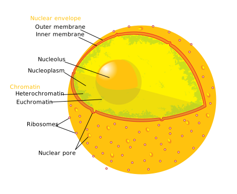
The nucleus is the largest structure in the cell, about 6 micrometres across, and it holds the DNA. Stretched end to end, the DNA in one human nucleus is about two metres long, packed into a space a millionth of that. Chapter 2.7 covers how that packing is done without tangling.
The nuclear envelope is a double membrane. Two complete bilayers, one inside the other, with a gap between them. The outer one is continuous with the endoplasmic reticulum, which means the space between the two membranes is the same space as the inside of the ER.
Nuclear pores are large protein assemblies that punch through both membranes, and there are roughly 2,000 to 4,000 of them per nucleus. They are not simple holes. Small molecules pass freely, but anything above about 40 kilodaltons needs a signal sequence — a short stretch of amino acids that acts as a passport — and a carrier protein to escort it through. This checkpoint is how the cell controls which transcription factors reach the DNA, and therefore which genes get switched on. Chapter 2.4 uses it constantly.
Chromatin is DNA wound around proteins called histones. It exists in two states: loosely packed, where genes can be read, and tightly packed, where they cannot. Which regions are which is a large part of what makes a liver cell different from a nerve cell despite identical DNA.
The nucleolus is the dark spot inside the nucleus, and it is not a compartment — it has no membrane. It is simply the region where ribosomal RNA is transcribed and ribosomes are assembled, dense enough with machinery to be visible. A cell making a lot of protein has a prominent nucleolus, which is one of the features a pathologist looks for when deciding whether a cell is cancerous.
When the nucleus fails: progeria. The inner face of the nuclear envelope is lined by a mesh of proteins called lamins that give it mechanical strength. A single-letter mutation in the LMNA gene produces a faulty lamin A that cannot be processed correctly, and the nuclear envelope becomes misshapen and fragile. The result is Hutchinson–Gilford progeria syndrome: children who appear to age at several times the normal rate, with hair loss, joint stiffness, and severe atherosclerosis, and who die typically in their early to mid teens of heart attack or stroke. It affects about one birth in four million. It is a stark demonstration that the nucleus is a structural organ, not just a container.
Ribosomes: where protein is built

A ribosome reads messenger RNA and builds the protein it specifies, joining amino acids at up to about twenty per second. A single cell may hold millions of them. Some float free in the cytosol, making proteins that stay inside the cell; others are stuck to the endoplasmic reticulum, making proteins destined for membranes or for export. Which one a ribosome becomes is decided by the protein it happens to start building — a signal sequence at the front of certain proteins causes the whole ribosome to be dragged to the ER while it works.
Ribosomes are measured by how fast they sediment in a centrifuge, in Svedberg units, written S. Your ribosomes are 80S, made of a 60S and a 40S subunit. Bacterial ribosomes are 70S, made of 50S and 30S. The numbers do not add up arithmetically because sedimentation depends on shape as well as mass.
That difference is a large part of your medicine cabinet. A drug that jams a 70S ribosome and ignores an 80S one will stop a bacterium from making proteins while leaving your cells untouched. Tetracyclines and aminoglycosides block the 30S subunit; macrolides such as azithromycin, and clindamycin, block the 50S. This is why these antibiotics can be given safely at doses that are lethal to bacteria — Chapter 22.6 covers the classes.
There is a catch, and it is a good illustration of how one fact explains a side effect. Your mitochondria have their own ribosomes, and they are 70S-like, because mitochondria descend from bacteria. So ribosome-targeting antibiotics do reach a bacterial-type target inside you. This is part of why aminoglycosides such as gentamicin can permanently damage the hair cells of the inner ear and cause irreversible deafness, and it is why they are used carefully and with blood level monitoring.
The endomembrane system: manufacture, packaging, dispatch

Rough endoplasmic reticulum is a stack of flattened membrane sheets continuous with the nuclear envelope, covered with ribosomes — the "rough" is those ribosomes seen under an electron microscope. Proteins built here are pushed through the membrane as they are made, ending up inside the ER's internal space. There they fold, get their first sugar chains attached, and are inspected. Misfolded proteins are held back and eventually destroyed, which is the quality control that traps the faulty CFTR of cystic fibrosis before it ever reaches the surface (Chapter 1.3).
Cells that export a lot of protein are packed with rough ER. A plasma cell pumping out antibodies is almost solid with it.
When the ER is overwhelmed — too many misfolded proteins accumulating — the cell triggers the unfolded protein response: it slows down all protein production, makes more chaperones, and steps up destruction of the backlog. If that fails to clear it, the cell kills itself. This is now understood to be part of how beta cells die in type 2 diabetes, where they have been forced to overproduce insulin for years.
Smooth endoplasmic reticulum has no ribosomes and does completely different jobs, which vary by tissue.
In the liver, it is the detoxification plant. A family of enzymes called cytochrome P450 sits in its membrane and chemically modifies foreign molecules — drugs, alcohol, toxins, and also your own steroid hormones — usually making them more water-soluble so the kidney can excrete them. This is where most drug metabolism happens, and it is where most drug interactions come from: two drugs handled by the same P450 enzyme compete, so one raises the other's blood level, sometimes into the toxic range. It is also why grapefruit juice carries a warning on many medicines — it contains compounds that inhibit one particular P450 enzyme, CYP3A4, so drugs handled by that enzyme accumulate. With some statins and some heart drugs this is enough to cause real harm. Chapter 22.13 lists the combinations that matter.
In muscle, the smooth ER is specialised into the sarcoplasmic reticulum, which stores calcium and releases it on command to trigger contraction (Chapter 6.1). In the testes, ovaries and adrenal glands, it is where steroid hormones are made.
The Golgi apparatus is a stack of flattened discs, named after Camillo Golgi, who first saw it in 1898 using a silver staining method he invented. It is the sorting office. Vesicles arrive from the ER at one face, move through the stack, and leave from the other. As they pass, sugar chains are trimmed and rebuilt, and address tags are attached that determine where each protein goes next.
The best-understood tag is a sugar called mannose-6-phosphate, which marks a protein for delivery to a lysosome. Break the enzyme that attaches that tag and you get I-cell disease, in which lysosomal enzymes are made perfectly but never delivered — they are secreted out of the cell instead, while the lysosomes fill with undigested material. The affected children have severe skeletal and developmental problems and usually do not survive childhood. It is an unusually clean demonstration that a cell's addressing system is as essential as its factories.
Lysosomes: the recycling plant
A lysosome is a membrane bubble containing about sixty different digestive enzymes, held at an internal pH of around 4.5 to 5 by a proton pump in its membrane. The name means "splitting body". It digests worn-out organelles, engulfed bacteria, and material brought in from outside.
The acid is the safety mechanism. Those enzymes only work at low pH, so if a lysosome leaks, its contents meet the neutral cytosol and largely stop working. The cell is protected by chemistry rather than by containment alone.
Lysosomal storage diseases are what happen when one of those sixty enzymes is missing. Its particular substrate is never broken down, so it accumulates inside lysosomes year after year until the cell can no longer function. There are more than fifty such diseases, each named for the missing enzyme's substrate, and they are individually rare but collectively not.
Tay–Sachs disease is the clearest example. The missing enzyme is hexosaminidase A; the substrate that accumulates is a lipid called GM2 ganglioside, found mostly in neurons. An affected infant appears normal at birth, meets early milestones, and then between three and six months of age begins to lose them — loss of head control, then sitting, then vision and hearing, with seizures, and death usually before age five. The diagnostic sign is a cherry-red spot in the back of the eye: the retina is thickened and pale with stored lipid everywhere except at the very centre, where the retina is thin, so the normal red of the blood supply shows through as a bright spot against pale surroundings.
Some of these diseases can now be treated by enzyme replacement therapy — infusing the missing enzyme, manufactured in cell culture and carrying the mannose-6-phosphate tag so the body's own machinery delivers it to lysosomes. It works for Gaucher disease and Fabry disease, among others. It does not work for Tay–Sachs, because infused enzyme cannot cross from blood into the brain, which is exactly where it is needed. That barrier is Chapter 11.10.
Peroxisomes: the chemical hazards unit
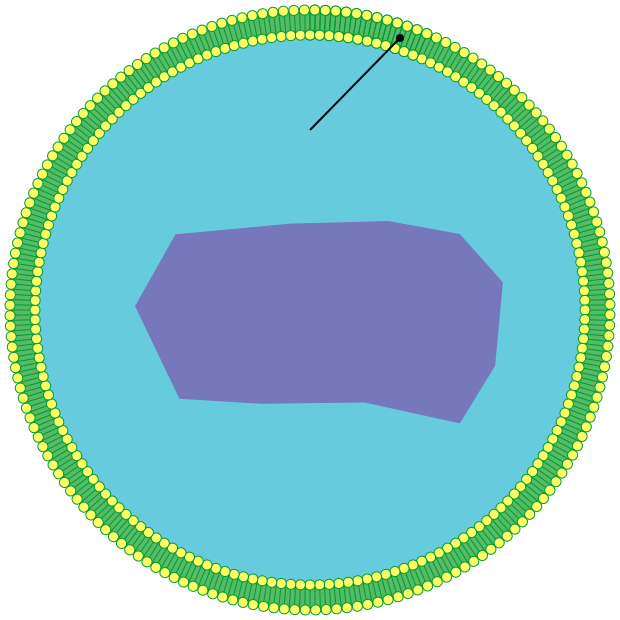
Peroxisomes handle reactions that generate hydrogen peroxide, H₂O₂ — a molecule that damages DNA and proteins on contact. They break down very long fatty acids that mitochondria cannot handle, and they detoxify various substances including a substantial share of the alcohol you drink.
The trick is that they contain their own antidote. Catalase, one of the fastest enzymes known, immediately splits hydrogen peroxide into water and oxygen. So the peroxide is made and destroyed inside a sealed sac and never meets the rest of the cell. This is also the reason hydrogen peroxide fizzes on a cut: catalase released from damaged cells is tearing it apart, and the bubbles are oxygen.
Peroxisomal diseases include Zellweger syndrome, where peroxisomes are not assembled at all and the newborn has severe brain, liver and kidney abnormalities, and X-linked adrenoleukodystrophy, where very long fatty acids accumulate and destroy the insulating sheath around nerve fibres. The second of these is the disease in the film Lorenzo's Oil, and the real story is more instructive than the film: the oil developed by the child's parents does lower the fatty acid level in blood, but trials showed it does not reverse existing neurological damage, though it may delay onset in boys treated before symptoms begin.
Mitochondria: the power stations, and the bacteria you are descended from

Mitochondria are where oxygen is used and where most of your ATP is made — Chapter 1.6 is entirely about the process. Each is about 1 to 2 micrometres long, roughly bacterium-sized. Numbers vary enormously with how much energy a cell needs: a red blood cell has none at all, a skin cell has a few hundred, a liver cell one to two thousand, and a heart muscle cell around five thousand, filling something like 30 to 40 percent of its volume. That single fact tells you what heart muscle is for and why it dies so fast when its blood supply is cut.
The inner membrane is folded into shelves called cristae, and the folding is not decoration. The ATP-making machinery is embedded in that membrane, so more membrane area means more output. In tissues with high demand the cristae are packed so densely that the interior is almost all membrane.
Mitochondria carry their own DNA. In humans it is a circular loop of 16,569 base pairs carrying 37 genes — a tiny genome compared with the roughly 20,000 genes in the nucleus, but essential ones. They also carry their own ribosomes, of the bacterial 70S type, and they divide by splitting in two rather than being built by the cell.
All of that is evidence for a specific historical event. Lynn Margulis argued in 1967, against considerable resistance, that mitochondria are the descendants of free-living bacteria that were engulfed by an ancestral cell and never digested — an arrangement called endosymbiosis, living together on the inside. The evidence is now overwhelming: the double membrane (the outer one being the host's engulfing membrane, the inner one the bacterium's own), the circular DNA, the bacterial ribosomes, the independent division, and the genetic sequence, which places mitochondria firmly among a specific group of bacteria. Chloroplasts in plants have the same origin from photosynthetic bacteria. Chapter 1.9 covers what this event made possible.
Mitochondrial DNA is inherited only from your mother. The egg contributes the cytoplasm and all its mitochondria; the sperm's few mitochondria are in the tail region and are destroyed after fertilisation. Three consequences follow.
Mitochondrial diseases pass only down the female line. An affected father has no affected children through this route; an affected mother may pass it to all of them. Leber's hereditary optic neuropathy, which causes sudden painless loss of central vision usually in young adult men, and MELAS, which causes stroke-like episodes in young people, are both mitochondrial.
The tissues that fail are the hungry ones — brain, heart, skeletal muscle, eye — because a partial energy shortfall shows up first wherever the demand is highest.
And mitochondrial DNA is a clock for tracing ancestry, since it does not get shuffled by sexual reproduction and changes only by mutation. This is where the idea of "mitochondrial Eve" comes from, and Chapter 3.5 explains what that phrase does and does not mean.
The cytoskeleton: shape, movement, and transport tracks
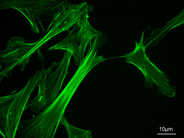
Three filament systems, of three different thicknesses, do three different jobs.
Microfilaments, about 7 nanometres across, are made of actin. They sit just under the cell membrane, control cell shape, and pull the cell into new shapes. Together with myosin they generate every contraction in your body — in muscle as an ordered array (Chapter 6.1), and in every other cell as the machinery that pinches a dividing cell in two and that lets a white blood cell crawl through tissue toward an infection.
Intermediate filaments, about 10 nanometres, are the pure tension-bearers. Keratins in skin cells, neurofilaments in nerve cells, and the lamins lining the nuclear envelope are all in this family. When they fail, tissues tear. In epidermolysis bullosa, faulty keratin means the skin blisters and sloughs off from ordinary friction.
Microtubules, about 25 nanometres, are hollow tubes of a protein called tubulin. They are the cell's railway. Motor proteins — kinesin walking one way, dynein the other — carry vesicles and organelles along them, hand over hand, one step per ATP. In a motor neuron running a metre down your leg, this is the only way anything gets from the cell body to the far end, and the trip takes days.
Microtubules also build the spindle that separates chromosomes during cell division (Chapter 1.7), and this makes them one of the most important drug targets in medicine.
- Taxanes, such as paclitaxel, originally from the Pacific yew tree, lock microtubules so they cannot come apart. The spindle freezes, division cannot complete, and the cell dies. Used in breast, ovarian and lung cancer.
- Vinca alkaloids, such as vincristine, from the Madagascar periwinkle, do the opposite — they prevent microtubules from assembling. Same outcome, opposite mechanism.
- Colchicine, from the autumn crocus and in use for gout since ancient Egypt, also blocks microtubule assembly. In gout it works by stopping white blood cells from crawling into the inflamed joint, since crawling needs a working cytoskeleton.
The reason all of these cause hair loss, nausea and low blood counts is now obvious: they hit every rapidly dividing cell, and hair follicles, gut lining and bone marrow are the fastest-dividing normal tissues you have. Chapter 19.7 covers the trade-off in full.
Centrioles are short barrels of microtubules that sit near the nucleus in a region called the centrosome and organise the spindle. Cilia and flagella are built from the same tubulin machinery: a flagellum is the tail that propels a sperm cell, and cilia are the hair-like projections that line your airways and sweep mucus upward at about one centimetre a minute.
When cilia do not work: primary ciliary dyskinesia. The affected person has chronic sinus and chest infections, because mucus is never cleared, and men are usually infertile, because sperm cannot swim. And about half of them have their internal organs mirror-imaged, with the heart on the right — a combination called Kartagener syndrome. The reason for that last part is genuinely elegant: in the very early embryo, a patch of cilia rotates to create a leftward flow of fluid, and that flow is what tells the body which side is left. Without working cilia there is no flow, no signal, and the sidedness is decided at random — so about half end up reversed. Chapter 4.5 covers it.
Getting rid of a single protein: the proteasome
Lysosomes handle bulk. Individual proteins that are damaged, misfolded or simply no longer needed are dealt with by the proteasome, a barrel-shaped protein complex in the cytosol that shreds them into short fragments.
Targets are marked for destruction by having a small protein called ubiquitin attached to them in a chain — a molecular label reading "destroy this". Aaron Ciechanover, Avram Hershko and Irwin Rose received the 2004 Nobel Prize in Chemistry for working out this system.
It is also a drug target. Bortezomib blocks the proteasome and is used in multiple myeloma, a cancer of antibody-producing plasma cells. It works because those cells are producing vast amounts of protein and therefore generating vast amounts of defective protein; block their disposal system and they choke on their own output faster than normal cells do.
Where this leaves you
Every organelle above is a place a disease can start, and the pattern repeats: a single missing enzyme, a single mis-addressed protein, a single failed filament, and the consequence appears in whichever tissue depends on it most.
It also means that when a doctor says a disease is "metabolic" or "genetic" or "degenerative", the real answer is usually a specific broken part in a specific compartment of a specific cell type. Most of the rest of this volume is about tracing that link in both directions — from the broken part forward to the symptom, and from the symptom back to the broken part.
What the next page fixes
Almost everything on this page cost ATP: the proton pump acidifying the lysosome, the motor proteins walking along microtubules, the vesicles budding and fusing, the sodium pump from Chapter 1.4 running continuously in the background. Chapter 1.6 follows a single glucose molecule from your mouth to the moment its energy is stored in ATP, through glycolysis, the Krebs cycle and the electron transport chain — and explains, finally, what the oxygen you breathe is actually for.