Appearance
4.2 — The Four Tissues
Your liver, your skin, your kidney and your gut look nothing alike. All four are built from the same four materials, in different proportions and arrangements — the way brick, timber, glass and steel build a house, a factory and a bridge that share no resemblance.
Knowing the four tissues is what lets you predict how a disease will behave, because a disease of one tissue type behaves similarly wherever it appears. A cancer of epithelium spreads one way, a cancer of connective tissue another, and knowing which you are dealing with is the first thing a pathologist establishes.
Tissue 1: epithelium — the linings

Epithelium covers every surface, inside and out. Your skin's outer layer, the lining of your gut, your airways, your bladder, your blood vessels, and the working cells of every gland are all epithelium.
Three universal features, and each explains something clinical.
Cells are packed tightly with almost no material between them, joined by specialised junctions. Some junctions seal the gap completely, so nothing can leak between cells and everything must pass through them — which is what makes an epithelium a genuine barrier and lets it control what crosses (Chapter 1.4). Others are mechanical rivets; others are pores connecting the insides of adjacent cells.
It sits on a basement membrane — a thin sheet of secreted material separating it from the tissue beneath. This sheet is the single most important structure in cancer staging. A cancer confined above it is in situ, cannot spread, and is cured by removing it. A cancer that has broken through it is invasive and can reach blood and lymph vessels. "Has it broken the basement membrane" is the question that separates a curable lesion from a potentially lethal one, and the whole point of cervical and breast screening is to catch cancers before they do (Chapter 19.4).
It has no blood vessels of its own. Oxygen and nutrients diffuse up from the tissue below. This limits how thick an epithelium can be, and it is why the outermost layers of skin are dead — they are too far from the supply.
Naming is two words: layers, then shape.
Simple squamous — one layer of flat cells. Thin enough to allow diffusion, so it lines every surface where exchange happens: the alveoli of the lungs, the walls of capillaries, the lining of blood vessels.
Simple cuboidal — one layer of box-shaped cells. Kidney tubules, glandular ducts. Cells thick enough to contain transport machinery.
Simple columnar — one layer of tall cells, often with microvilli on top to multiply surface area and often with mucus-producing goblet cells among them. This lines your stomach and intestines, and it is built for absorption and secretion.
Pseudostratified columnar — one layer that looks like several because the nuclei sit at different heights. Lines the airways, usually with cilia on top and goblet cells producing mucus. This is the escalator that clears your lungs: goblet cells make the mucus, cilia beat it upward at about a centimetre a minute, and you swallow it without noticing. Cigarette smoke paralyses the cilia within minutes and eventually destroys them, which is exactly why smokers cough — the escalator is broken and coughing is the only remaining way to clear it, and it is why the cough is worst in the morning after a night of accumulation.
Stratified squamous — many layers, flat at the top, built for abrasion. Skin, mouth, oesophagus, vagina. On skin the top layers are filled with keratin and dead; on wet surfaces they are not.
Transitional — many layers that change shape as the tissue stretches. Found only in the urinary tract, and it lets your bladder expand from empty to about 500 ml without tearing and without letting urine leak into the tissue.
Glands are epithelium too. Exocrine glands secrete through a duct onto a surface — sweat, saliva, digestive enzymes, milk. Endocrine glands have no duct and secrete into the blood — the hormones of Part 12.
And 80 to 90 percent of human cancers arise from epithelium, because it is the tissue that divides constantly and the tissue most exposed to the outside world. A cancer of epithelium is called a carcinoma.
Tissue 2: connective tissue — the framework
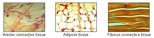
The defining feature is the reverse of epithelium: cells are sparse and the material between them dominates. That material — the extracellular matrix — is what gives each connective tissue its properties.
Three fibre types:
Collagen is the most abundant protein in your body, around 25 to 30 percent of all your protein. It is a rope of three intertwined chains, and it resists pulling — a collagen fibre has tensile strength comparable to steel wire of the same diameter. There are at least 28 types; type I is in skin, bone and tendon, type II in cartilage, type IV in basement membranes.
Making collagen requires vitamin C. The three chains only lock together properly after specific amino acids are chemically modified by an enzyme that needs vitamin C as a cofactor. Without it, collagen is unstable, and this is exactly what scurvy is: bleeding gums, teeth falling out, old wounds reopening, capillaries breaking, and eventual death. Old scars reopening is the giveaway sign, and it makes complete sense once you know that a scar is collagen that must be continuously renewed. Vitamin C reverses it in days.
Elastin allows recoil. Skin, lung and artery walls contain it, and its gradual loss with age is why skin no longer springs back when pinched.
Reticular fibres form fine supporting networks in organs like the liver and spleen.
The types of connective tissue:
Loose areolar — the packing material that sits under every epithelium and around every organ. It holds blood vessels, nerves and immune cells, and it is where most inflammation happens.
Adipose — fat. Cells almost entirely filled with a single lipid droplet. Energy store, insulation, mechanical cushion — and an endocrine organ in its own right, secreting leptin, adiponectin and inflammatory signals, which is the reason obesity is a systemic inflammatory condition rather than just stored weight (Chapter 18.8).
Dense regular — collagen fibres all in parallel. Tendon (muscle to bone) and ligament (bone to bone). Enormously strong along the line of pull and weak across it, which is why tendons and ligaments tear when force comes from an unexpected direction. Their blood supply is poor, so they heal slowly — a torn ligament can take months, against days for a cut in well-supplied skin.
Dense irregular — collagen in all directions. The deep layer of skin, and the capsules around organs. Strong in every direction rather than very strong in one.
Cartilage — cells in a firm gel matrix, with no blood vessels at all. Nutrients diffuse in from the surface, which is why cartilage heals extremely poorly and why joint cartilage damage tends to be permanent (Chapter 5.8). Three kinds: hyaline on joint surfaces and in the airways, fibrocartilage in intervertebral discs and knee menisci, elastic in the ear and epiglottis.
Bone — matrix hardened with calcium phosphate crystals, and very much alive with its own blood supply (Chapter 5.1).
Blood — a connective tissue, which surprises everyone. It qualifies because it is cells suspended in an extensive extracellular matrix; the matrix is simply liquid plasma rather than fibres and gel (Chapter 7.1).
Cancers of connective tissue are called sarcomas. They are far rarer than carcinomas, tend to occur in younger people, and spread through the bloodstream rather than the lymphatics.
And several inherited diseases are collagen faults. Osteogenesis imperfecta — brittle bone disease — is a type I collagen defect, giving bones that fracture from minimal force and often blue sclerae, because the thin collagen of the white of the eye lets the dark layer beneath show through. Ehlers–Danlos syndromes cause hypermobile joints and fragile skin, and one form causes arteries to rupture. Marfan syndrome affects fibrillin, a component of elastic fibres, giving tall stature, long fingers, lens dislocation and — the dangerous part — a weak aortic wall that can tear (Chapter 21.5).
Tissue 3: muscle
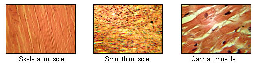
All three contract using the same actin and myosin machinery (Chapter 6.1). What differs is arrangement and control.
Skeletal muscle. Long cylindrical fibres with visible stripes, formed by many cells fusing so that each fibre has many nuclei. Under voluntary control. Contracts fast and fatigues. Attached to bone by tendons.
Cardiac muscle. Striped like skeletal muscle but branched, with one or two nuclei per cell, and joined end to end by structures called intercalated discs. Those discs contain electrical connections that let current pass directly from cell to cell, so the heart contracts as a single coordinated unit rather than fibre by fibre — which is exactly what a pump needs and what a limb muscle must not have. It never fatigues, and it is under involuntary control (Chapter 7.2).
Smooth muscle. Spindle-shaped cells, no stripes, one nucleus. Lines every hollow organ: blood vessels, gut, airways, bladder, uterus. Involuntary, slow, and able to sustain contraction for long periods at very low energy cost. This is the tissue most drugs act on when they change blood pressure, relieve asthma, or treat gut cramps.
And this is where cardiac muscle's limitation from Chapter 1.7 matters most. Skeletal muscle has satellite cells that can repair damage. Cardiac muscle cells have essentially left the cell cycle permanently, so dead heart muscle is replaced by scar, not by muscle. Chapter 18.4.
Tissue 4: nervous tissue
Two cell populations, and the supporting one is far more numerous than most people assume.
Neurons carry electrical signals. A neuron has a cell body containing the nucleus, many dendrites receiving signals in, and one axon carrying the signal out — which may be over a metre long (Chapter 11.1).
Glia support them, and there are roughly as many glial cells as neurons in the human brain — the often-quoted ten-to-one ratio is not supported by careful counting. Four main types:
- Astrocytes regulate the chemical environment, supply neurons with fuel, and form part of the blood–brain barrier.
- Oligodendrocytes in the brain and spinal cord wrap axons in myelin, the fatty insulation that makes conduction fast. Multiple sclerosis is an attack on these cells (Chapter 20.5).
- Schwann cells do the same job in peripheral nerves, and unlike oligodendrocytes they support regeneration — which is why a cut peripheral nerve can recover and a cut spinal cord cannot.
- Microglia are the brain's immune cells.
Nervous tissue's defining limitation is that neurons in the central nervous system do not regenerate meaningfully. Damage is permanent, and recovery after a stroke or spinal injury comes from surviving circuits taking over function rather than from replacement.
Putting them together
Every organ is a combination, and the pattern is remarkably consistent. Take the small intestine from the inside out:
Epithelium lines the lumen, absorbing nutrients. Loose connective tissue beneath it carries blood vessels and lymphatics. Smooth muscle in two layers — one circular, one longitudinal — moves the contents along. Nervous tissue forms a network between the muscle layers that coordinates the movement. And connective tissue wraps the whole tube.
Skin is the same logic: stratified squamous epithelium on top, dense irregular connective tissue beneath, with smooth muscle attached to hair follicles, sensory nerve endings throughout, and adipose tissue below.
And this is why a pathologist can tell what a tumour is from what it looks like, and why the answer changes the treatment entirely.
What the next page fixes
You now know what the body is made of. The remaining question is how it got assembled — because nothing on this page explains how a single cell produces four tissue types in the right arrangement. Chapter 4.3 starts that story at fertilisation: what actually happens when a sperm meets an egg, how the resulting cell divides for the first week, and why the timing of implantation determines whether a pregnancy happens at all.