Appearance
8.2 — The Lungs and the Pleura
Your lungs contain around 300 to 500 million alveoli, giving a gas exchange surface of roughly 70 square metres — about the area of a tennis court, folded into a space the size of two loaves of bread. The barrier between air and blood is about 0.5 micrometres thick, thinner than a red blood cell, and across it you move about 250 ml of oxygen and 200 ml of carbon dioxide every minute at rest.
They also weigh almost nothing — around 1 kilogram for the pair, most of which is blood — and they float in water, which is why the technique of putting lung tissue in water is used in forensic pathology to determine whether a newborn ever breathed.
Shape and lobes
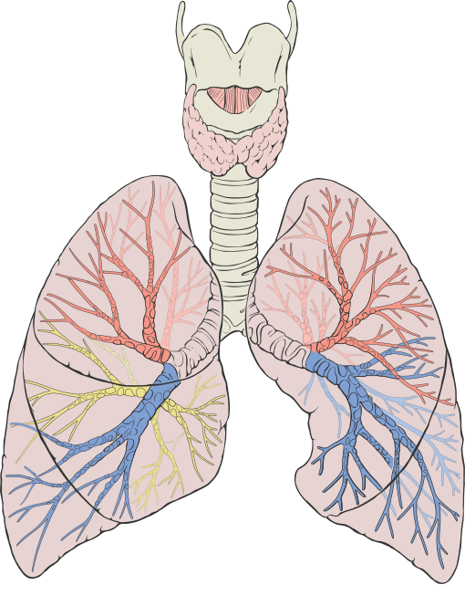
Right lung: three lobes — upper, middle and lower, separated by an oblique and a horizontal fissure.
Left lung: two lobes — upper and lower. The heart takes up the space where a middle lobe would be, leaving a notch in the front border of the left upper lobe called the cardiac notch. The tongue-shaped projection below it, the lingula, is the left lung's equivalent of a middle lobe.
So the right lung is about 10 percent larger than the left.
Each lobe divides into bronchopulmonary segments — 10 on the right and 8 to 10 on the left — each with its own segmental bronchus and artery, and each surrounded by connective tissue.
That segmental independence is surgically important. A single segment can be removed while leaving the rest of the lobe intact, which is increasingly done for small lung cancers instead of removing a whole lobe, preserving lung function.
And it is why pneumonia can be confined to a recognisable shape on a chest X-ray — the infection fills one segment or lobe and stops abruptly at its boundary.
The hilum is where the main bronchus, the pulmonary artery, the pulmonary veins, the lymphatics and the nerves enter and leave. Enlarged hilar lymph nodes on a chest X-ray are a significant finding, seen in tuberculosis, sarcoidosis and lung cancer.
The pleura
Two membranes: the visceral pleura covering the lung surface, and the parietal pleura lining the inside of the chest wall, the diaphragm and the mediastinum. They are continuous with each other at the hilum, like a fist pushed into a partly inflated balloon.
Between them is the pleural space, containing only about 10 to 20 ml of fluid — a film a few micrometres thick.
That film does two things, and the second is the one people miss.
It lubricates, so the lung slides against the chest wall with almost no friction through 20,000 breaths a day.
And it couples the lung to the chest wall by surface tension. Two wet glass slides slide against each other freely but cannot easily be pulled apart. The lung is held to the chest wall the same way — so when the chest expands, the lung has no choice but to expand with it, even though nothing physically attaches them.
This is the whole mechanism of breathing, and Chapter 8.3 develops it.
The pressure in the pleural space is negative, about −5 cmH₂O at rest, falling to about −8 during inspiration. It is negative because the lung's elastic recoil pulls inward while the chest wall's elasticity pulls outward, and the two opposing pulls create a suction between them.
Two nerve supplies, and the difference is diagnostic.
The visceral pleura has no pain fibres. Lung tissue itself is insensitive — which is why lung cancer can grow to a considerable size with no pain at all, and is so often diagnosed late.
The parietal pleura is richly supplied with pain fibres from the intercostal nerves. So chest pain from a lung problem only occurs once the process has reached the parietal pleura.
Pleuritic pain is therefore characteristic and easy to recognise: sharp, localised, and worse on deep breathing, coughing or moving. It is the sound of two inflamed surfaces rubbing, and a rubbing sound is sometimes audible with a stethoscope.
And the diaphragmatic parietal pleura is supplied by the phrenic nerve, so irritation there refers to the shoulder tip (Chapter 6.4).
Pleural problems
Pneumothorax — air in the pleural space. The surface-tension coupling is broken, so the lung's elastic recoil pulls it inward and it collapses.
Primary spontaneous pneumothorax occurs without underlying lung disease. The typical patient is a tall, thin, young man who smokes, and the mechanism is a small bleb at the lung apex rupturing. The height association is genuinely mechanical: pleural pressure is more negative at the apex than at the base in an upright person, and the taller the chest, the greater that gradient, so the apical alveoli are more distended.
Secondary pneumothorax occurs in existing lung disease — COPD, cystic fibrosis, asthma — and is more dangerous because there is less reserve.
Tension pneumothorax — the emergency version described in Chapter 5.4. Air enters through a one-way flap and cannot escape, pressure rises, the mediastinum shifts and venous return is obstructed. Treated immediately with a needle, before imaging.
Pleural effusion — fluid in the space. Can be a litre or more.
The key distinction is between a transudate and an exudate, and it is made using Light's criteria, comparing protein and enzyme levels in the fluid with those in blood.
A transudate is a filtration problem — the pleural membranes are normal, and fluid is being pushed out or not pulled back by the Starling forces of Chapter 7.5. Causes: heart failure, low albumin, liver failure.
An exudate means the membranes are abnormal and leaking. Causes: infection, cancer, pulmonary embolism, inflammation.
That single test therefore separates "the plumbing is wrong somewhere else" from "something is wrong with the pleura itself", and it directs everything that follows.
Empyema is pus in the pleural space, usually complicating pneumonia. It needs drainage as well as antibiotics, because antibiotics penetrate a walled-off collection of pus poorly.
A chest drain is inserted in the "safe triangle" in the armpit region — bounded by the edge of the pectoralis major in front, the latissimus dorsi behind, and a line at the level of the nipple below. The triangle exists because it avoids the muscle bulk, the long thoracic nerve, and the diaphragm.
And the drain is inserted immediately above a rib, never below one, because the intercostal vessels and nerve run in a groove on the underside of each rib. Going in just above the lower rib of the space keeps well clear of them.
The alveoli
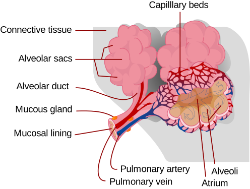
Each alveolus is about 0.2 to 0.3 millimetres across, and they are wrapped so densely in capillaries that blood forms almost a continuous sheet around them.
The barrier consists of three layers: the alveolar cell, a fused basement membrane, and the capillary endothelial cell. Total thickness 0.2 to 0.6 micrometres.
Two cell types line the alveolus:
Type I cells — extremely flat, covering about 95 percent of the surface. They are the barrier, and being thin is their entire job.
Type II cells — cuboidal, about 5 percent of the surface but 60 percent of the cells by number. They produce surfactant, and they are the stem cells that regenerate the alveolar lining after injury.
Alveolar macrophages patrol the surface, swallowing inhaled particles and bacteria. They are why the deep lung is essentially sterile despite processing 10,000 litres of unfiltered air a day.
In heart failure, macrophages that have engulfed red cells leaking into the alveoli appear in the sputum, and they were historically called "heart failure cells".
Surfactant
A tiny bubble is harder to keep open than a large one, and this is not intuitive. Laplace's law for a sphere with a liquid lining gives:
P = \frac{2T}{r}
Pressure needed is inversely proportional to radius. So a small alveolus needs more pressure to stay open than a large one.
Which creates a serious problem. If small and large alveoli are connected — and they are, through the airways — the small one should empty into the large one, because it has the higher pressure. The lung should collapse into a few enormous sacs, destroying its surface area.
Surfactant solves it. It is a mixture of phospholipids and proteins produced by type II cells, which spreads as a film over the liquid lining and reduces surface tension.
And it does so in a radius-dependent way. As an alveolus shrinks, the surfactant molecules are pushed closer together and the film becomes more concentrated, so surface tension falls further. In a large alveolus, the film is spread thin and tension is higher.
So surfactant lowers T more in small alveoli than in large ones, which cancels out the effect of the smaller r, and alveoli of different sizes coexist stably. It is a self-correcting mechanism built into a chemical film.
Surfactant also has two other effects. It reduces the work of breathing substantially — without it, the effort required to inflate the lungs would be several times greater. And it keeps alveoli dry, because surface tension otherwise pulls fluid out of the capillaries into the air spaces.
Surfactant production begins around 24 weeks of gestation and reaches adequate levels around 34 to 36 weeks.
This is why premature babies develop respiratory distress syndrome. Without enough surfactant, alveoli collapse at the end of each breath and must be reinflated from scratch, which takes enormous effort. The baby tires, and the collapsed lung leaks fluid and protein.
Two treatments transformed this, and both come straight from the physiology.
Antenatal corticosteroids given to the mother 24 to 48 hours before a preterm delivery accelerate surfactant production. This single intervention reduces neonatal death by around 30 percent, and it is one of the most cost-effective treatments in all of medicine.
Artificial surfactant delivered directly into the lungs after birth. Introduced in the 1990s, and it cut mortality from respiratory distress syndrome by around 40 percent.
Survival at 28 weeks was roughly 20 percent in 1970 and is over 90 percent in well-resourced units today, and these two treatments are a large part of the reason.
Blood supply — two systems
The lungs have two entirely separate blood supplies, which is unusual.
The pulmonary circulation carries the entire cardiac output at low pressure, for gas exchange. This blood is not for the lung; it is being processed by the lung.
The bronchial circulation comes from the aorta at systemic pressure and supplies the lung tissue itself with oxygenated blood — the airways down to the terminal bronchioles, and the connective tissue.
This dual supply is why a pulmonary embolism does not always kill the lung tissue behind it. The bronchial arteries can keep it alive. Lung infarction after an embolus occurs in only a minority of cases, mostly where the bronchial supply is also compromised.
And bronchial arteries are the source of massive haemoptysis — coughing up large volumes of blood — because they are at systemic pressure. Treatment is often to block them with a catheter rather than to operate.
The lung's other jobs
Not only gas exchange.
It is a filter. Small clots, fat globules and debris are trapped in the pulmonary capillaries and broken down, rather than reaching the brain. The lung protects the arterial circulation from everything the venous circulation collects — which is exactly what a patent foramen ovale bypasses (Chapter 7.9).
It is a metabolic organ. Angiotensin-converting enzyme is concentrated in the pulmonary capillary endothelium, so the lung is where angiotensin I becomes angiotensin II (Chapter 7.6). It also inactivates several other circulating substances.
It is a blood reservoir, holding about 500 ml, which can be shifted to the systemic circulation when needed.
It contributes to acid–base balance by controlling carbon dioxide, and it does so within minutes where the kidney takes days (Chapter 10.4).
And it is a heat and water loss route — you lose around 300 to 400 ml of water a day simply by exhaling saturated air.
What the next page fixes
The lungs contain no muscle and cannot inflate themselves. Chapter 8.3 covers how they are inflated from outside — the pressures involved, why the coupling described on this page is what makes it work, and what "work of breathing" actually means when someone is struggling to breathe.