Appearance
9.2 — The Stomach
Your stomach contains hydrochloric acid at around pH 1.5 to 2 — strong enough to dissolve a razor blade in a week, and about a hundred thousand times more acidic than your blood. It secretes about 2 litres of it a day.
And it does not digest itself, which is not because the wall is chemically resistant — it is not — but because it maintains an active, continuously renewed defence. Understanding that defence explains almost every stomach disease and every stomach drug.
Structure
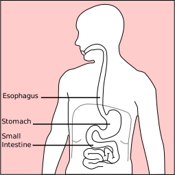
A J-shaped bag holding about 50 ml when empty and comfortably 1 to 1.5 litres after a meal, distensible to around 4 litres.
Five regions:
- Cardia — where the oesophagus enters.
- Fundus — the dome above the entrance, which collects swallowed gas. This is why the gas bubble sits at the top and why you belch when upright.
- Body — the largest part, and where most acid is produced.
- Antrum — the lower portion, which does the mechanical grinding.
- Pylorus — the muscular outlet valve.
Rugae are the folds of the inner lining, which flatten as the stomach fills, allowing expansion without stretching the tissue itself.
The muscle wall has three layers rather than the usual two — an extra oblique layer inside the circular and longitudinal ones. This is what allows the churning motion, which is a genuine grinding rather than simple squeezing.
What the stomach actually does
Store. It lets you eat a meal in minutes and release it over hours. Without a stomach you can survive and must eat small amounts frequently.
Mechanical breakdown. Contractions of about three per minute sweep from body to pylorus. The pylorus is almost closed, so most contents are squirted back — retropulsion — and this back-and-forth is what grinds the food. Only particles under about 2 millimetres pass through.
Chemical breakdown. Acid and pepsin begin protein digestion.
Kill microbes. The acid barrier is one of the body's most important defences, and its loss matters. People on long-term acid suppression have a measurably increased risk of certain gut infections, particularly Clostridioides difficile and Salmonella, precisely because the barrier is down.
Absorb almost nothing. The stomach absorbs essentially no nutrients. It absorbs alcohol, aspirin, and some water. This is why alcohol on an empty stomach affects you faster — with food present, the stomach empties more slowly and less alcohol is absorbed directly.
Make intrinsic factor — and this is the one function that is absolutely essential. Intrinsic factor is required for vitamin B12 absorption in the terminal ileum (Chapter 7.1). Everything else the stomach does can be compensated for; this cannot. Anyone who has had their stomach removed needs lifelong B12 injections, and without them develops irreversible neurological damage.
The cells
Parietal cells — produce hydrochloric acid and intrinsic factor.
The acid-making mechanism is worth following, because it is the target of the most-prescribed drug class in the world.
Inside the cell, carbonic anhydrase converts CO₂ and water into hydrogen and bicarbonate ions (Chapter 8.4). The hydrogen ion is then pumped into the stomach lumen by the H⁺/K⁺ ATPase — the "proton pump" — in exchange for potassium, using ATP. Chloride follows.
The pump can generate a concentration gradient of about three million to one, which is the steepest ion gradient in the human body.
And the bicarbonate produced goes the other way, into the blood. So after a meal, as acid production peaks, the blood leaving the stomach becomes transiently alkaline — the alkaline tide, briefly making the urine less acidic.
Chief cells — produce pepsinogen, the inactive precursor of pepsin.
It is secreted inactive deliberately. Active pepsin inside the cell would digest the cell. Acid converts pepsinogen to pepsin in the lumen, and pepsin then activates more pepsinogen — positive feedback (Chapter 4.7), so the process accelerates once started.
Pepsin works only below about pH 5, so it stops the moment the contents reach the alkaline duodenum. This is a neat safety design: the enzyme switches itself off by leaving the environment it needs.
G cells — in the antrum, producing gastrin, the hormone that stimulates acid production.
Mucous cells — produce the mucus and bicarbonate that protect the lining.
Enterochromaffin-like cells — release histamine, which stimulates parietal cells.
The defence
Three layers stop the stomach digesting itself.
A mucus layer about 0.2 millimetres thick, physically separating acid from cells.
Bicarbonate secreted into that mucus, so that although the lumen is at pH 2, the surface of the cells beneath the mucus is at pH 7. There is a pH gradient of five units across a fraction of a millimetre.
Tight junctions and rapid turnover. The lining cells are joined tightly so acid cannot leak between them, and the entire surface epithelium is replaced every 3 to 6 days.
All three depend on good blood flow, which supplies the bicarbonate and the energy for renewal, and prostaglandins maintain that blood flow and stimulate mucus and bicarbonate production.
That last sentence is the whole explanation of why NSAIDs cause ulcers. Aspirin and ibuprofen block cyclooxygenase, which makes prostaglandins (Chapter 1.3). Less prostaglandin means less mucus, less bicarbonate and less blood flow — so the defence fails while the acid continues.
And it is why NSAID damage happens even when the drug is not swallowed. An NSAID given by injection or suppository still causes stomach damage, because the mechanism is systemic prostaglandin blockade rather than local irritation.
Controlling acid production
Three phases, which together explain why hunger, smell and stress all produce stomach symptoms.
Cephalic phase (about 30 percent) — triggered by the sight, smell, taste and even thought of food, through the vagus nerve. Acid production begins before anything is eaten.
Gastric phase (about 60 percent) — triggered by distension and by the presence of protein, acting through gastrin.
Intestinal phase (about 10 percent) — initially stimulatory, then predominantly inhibitory as acidic chyme in the duodenum triggers hormones that shut acid production down.
Three stimulators converge on the parietal cell: acetylcholine from the vagus, gastrin from G cells, and histamine from ECL cells.
And this arrangement explains the two drug classes exactly.
H2 blockers (ranitidine, famotidine) block only the histamine input. They reduce acid by around 70 percent, because the other two routes remain.
Proton pump inhibitors (omeprazole, lansoprazole) block the pump itself — the final common step. They reduce acid by over 90 percent regardless of which signal is arriving. This is why they are so much more effective, and it is a good illustration of a general principle in pharmacology: blocking a final common pathway beats blocking one of several inputs.
PPIs also have a quirk worth knowing. They only inhibit pumps that are actively secreting, so they work best taken 30 to 60 minutes before a meal, when the pumps are being switched on. Taken with or after food they are substantially less effective, and this is one of the commonest reasons a PPI appears not to work.
Gastric emptying
Liquids empty fast, solids slowly. A meal takes 2 to 4 hours; a fatty meal considerably longer.
Fat is the most powerful brake, acting through hormones released by the duodenum — which makes sense, because fat is the slowest thing to digest and the duodenum needs time.
This is why a fatty meal "sits heavily" and why fat slows the absorption of anything taken with it.
Gastroparesis — delayed emptying — causes nausea, early fullness, bloating and vomiting of food eaten hours earlier. Diabetes is the commonest cause, through autonomic nerve damage, and it makes blood sugar control very difficult because the timing of absorption becomes unpredictable relative to insulin.
Dumping syndrome is the opposite, usually after stomach surgery. Rapid delivery of concentrated food into the small intestine pulls water in osmotically, causing cramping, diarrhoea, sweating and palpitations 15 to 30 minutes after eating, and sometimes a reactive low blood sugar an hour or two later.
Peptic ulcer disease, and one of medicine's best stories
A peptic ulcer is a break through the full thickness of the mucosa, in the stomach or the duodenum.
For most of the twentieth century, ulcers were believed to be caused by stress and spicy food, and treated with bland diets, antacids, and — when severe — surgery to cut the vagus nerve or remove part of the stomach. The dogma was that no bacterium could survive in stomach acid.
In 1982, Barry Marshall and Robin Warren in Perth, Australia, found a spiral bacterium in the stomach lining of ulcer patients and proposed it as the cause. They were largely ignored, and their paper was rejected.
In 1984, Marshall drank a culture of it. He developed gastritis within days, demonstrated the bacteria on biopsy, and cured himself with antibiotics.
They received the Nobel Prize in 2005. Helicobacter pylori is now known to cause the great majority of peptic ulcers, and ulcer disease has been transformed from a chronic condition managed for life into an infection cured in a week.
How the bacterium survives is genuinely clever: it produces urease, an enzyme that splits urea into ammonia and carbon dioxide. The ammonia is alkaline and neutralises acid in a cloud around the organism, and the bacterium burrows into the mucus layer where the pH is already near neutral.
And that same enzyme is how it is detected. The urea breath test has the patient swallow urea labelled with a traceable carbon isotope. If H. pylori is present, its urease splits the urea and the labelled carbon appears in the breath as CO₂ within minutes. A diagnostic test built directly on the organism's survival mechanism.
About half the world's population carries it, mostly without symptoms. It causes:
- Duodenal ulcers — over 90 percent are H. pylori related.
- Gastric ulcers — around 70 to 80 percent.
- Gastric cancer — it is classified as a Group 1 carcinogen.
- MALT lymphoma — a lymphoma of the stomach lining that frequently regresses completely when the bacterium is eradicated, which is one of the very few cancers curable with antibiotics.
Treatment is "triple therapy" — a proton pump inhibitor plus two antibiotics for 7 to 14 days, with regimens varying by local resistance patterns.
The two causes of peptic ulcer are therefore H. pylori and NSAIDs, and between them they account for the great majority. Stress and diet are contributory at most.
Ulcer symptoms: burning upper abdominal pain. Duodenal ulcer pain is classically relieved by food and returns 2 to 3 hours later, often waking the person at night. Gastric ulcer pain is often worsened by eating.
Two complications matter.
Bleeding — the commonest, presenting as vomiting blood or "coffee grounds", or black tarry stool (melaena), which is black because haemoglobin has been digested during its passage through the gut. This is an emergency.
Perforation — the ulcer erodes right through the wall, spilling contents into the abdominal cavity. Sudden severe pain, a rigid board-like abdomen, and a person who lies completely still because any movement is agonising. Free gas is visible under the diaphragm on an upright chest X-ray. Surgical emergency.
Stomach cancer
Incidence has fallen dramatically over the last century in most of the world, and the reasons are instructive: refrigeration replaced salting and smoking as the main food preservation methods, and H. pylori prevalence has fallen.
It remains common in East Asia, where screening endoscopy programmes in Japan and South Korea detect it early and have substantially improved survival.
The problem elsewhere is late presentation, because early symptoms are vague — indigestion, mild discomfort, early fullness. The alarm symptoms from Chapter 9.1 are what trigger investigation, and applying them properly is the main lever available.
Vomiting
A coordinated reflex, not simply reverse peristalsis.
The sequence: a deep breath, the glottis closes, the soft palate rises to seal the nose, the abdominal muscles and diaphragm contract forcefully and squeeze the stomach between them, and the lower oesophageal sphincter relaxes.
The force comes from the abdominal muscles, not the stomach, which is why vomiting is exhausting and why it is impaired in people with weak abdominal muscles.
The vomiting centre in the brainstem receives inputs from five sources, and each has its own drug class — which is why the choice of anti-sickness drug depends on the cause.
- The chemoreceptor trigger zone, which sits outside the blood–brain barrier so it can sample the blood directly. Drugs, toxins and metabolic disturbances act here. Blocked by dopamine antagonists such as metoclopramide and by ondansetron.
- The vestibular system — motion sickness. Blocked by antihistamines and hyoscine, which is why travel sickness tablets are antihistamines.
- The gut, via vagal afferents responding to distension, irritation or toxins.
- Higher centres — sight, smell, memory, anxiety. This is why anticipatory nausea before chemotherapy is real and why it responds to benzodiazepines rather than to conventional antiemetics.
- Raised intracranial pressure, which causes vomiting characteristically without much nausea, often in the early morning, and with headache.
Prolonged vomiting causes a specific metabolic picture: loss of hydrogen and chloride causes a metabolic alkalosis with low chloride and low potassium, and it is corrected with saline and potassium (Chapter 10.4).
What the next page fixes
The stomach delivers a semi-liquid acidic mixture into the duodenum, where the real work happens. Chapter 9.3 covers the small intestine — 6 metres of tube with a surface area of a badminton court, where essentially everything you absorb is absorbed.