Appearance
1.7 — The Cell Cycle and Division
You replace roughly 330 billion cells every day. Most of them are blood cells and the lining of your gut, and the turnover rates are not gentle: the cells lining your small intestine last three to five days before being shed into the bowel and digested, the outer layer of your skin turns over in about four weeks, and a red blood cell lasts 120 days before being broken up in your spleen. Over a year you rebuild a substantial fraction of yourself.
Meanwhile the neurons in your brain and most of your heart muscle cells are the ones you were born with, and will not be replaced.
Both of those facts come from the same set of controls: a cell decides whether to divide, and the decision is made by a system of checkpoints that can be wrong in two directions. Fail to divide when you should, and a wound never closes and a bone never heals. Divide when you should not, and that is cancer. This page is about that decision.
The cycle
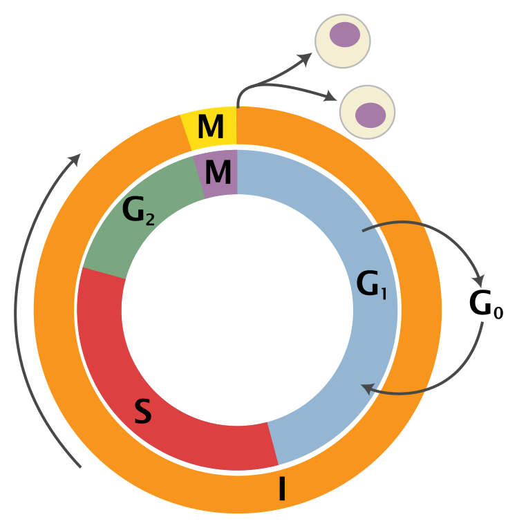
A dividing human cell takes roughly 24 hours to go round, though this varies enormously by tissue. The phases:
G1 (about 11 hours) — the gap after division. The cell grows, makes proteins, and produces more organelles. This is also where the decision to divide is actually taken.
S (about 8 hours) — synthesis. Every one of the cell's 46 chromosomes is copied, giving 92 DNA molecules held in 46 pairs. Chapter 2.2 covers how the copying works and how it avoids errors.
G2 (about 4 hours) — the second gap. The cell checks the copy, repairs what it can, and builds the machinery for division.
M (about 1 hour) — mitosis and cytokinesis: the chromosomes are separated and the cell splits in two.
G0 is not a phase of the cycle but an exit from it. A cell in G0 is alive and working but not preparing to divide. Your liver cells sit in G0 and will re-enter the cycle if part of the liver is removed — which is why a living donor can give away most of a liver lobe and both people regrow to near-normal volume within months. Your neurons and heart muscle cells enter G0 permanently. That is why a heart attack leaves a permanent scar of fibrous tissue rather than new muscle, and it is one of the central problems of cardiology.
Chromosomes, quickly
During most of the cycle the DNA is spread out as loose chromatin, because it has to be readable. Just before division it condenses into the compact X-shaped bodies everyone recognises as chromosomes, because a two-metre tangle cannot be sorted but 46 short rods can.
After the S phase, each chromosome consists of two identical sister chromatids joined at a waist called the centromere. That X shape is one chromosome carrying two copies, not two chromosomes.
You have 46 chromosomes in 23 pairs — one of each pair from your mother, one from your father. A cell with both members of every pair is diploid, written 2n. A cell with one of each is haploid, written n. All your body cells are diploid; only eggs and sperm are haploid.
Mitosis: making two identical cells
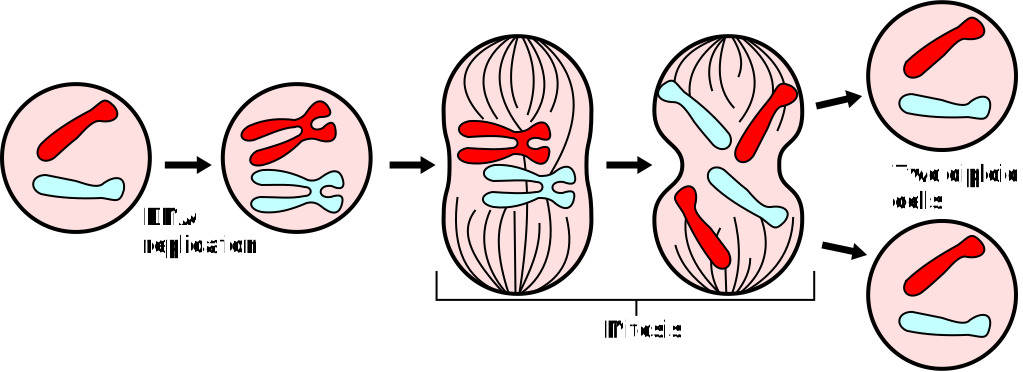
Prophase. Chromatin condenses into visible chromosomes, each already doubled into two sister chromatids. The nucleolus disappears. The centrosomes, each carrying a pair of centrioles, move to opposite ends of the cell and begin growing microtubules between them — the spindle of Chapter 1.5.
Prometaphase. The nuclear envelope breaks down. Spindle microtubules reach in and attach to a protein platform called the kinetochore, built on each chromatid's centromere. Each chromosome must end up attached to both poles — one chromatid to each side. Getting this right is the entire point of the exercise.
Metaphase. The chromosomes are pulled into a single line across the middle of the cell, each held in a tug-of-war balanced between the two poles. This is the arrangement in which chromosomes are photographed for a karyotype, the chromosome picture used to diagnose Down syndrome and similar conditions.
Anaphase. The link holding the sister chromatids together is cut, all at once, and the two sets are dragged to opposite poles. It takes only a few minutes. The all-at-once part matters: if one chromosome separated early, one daughter cell would get an extra copy and the other would be short one.
Telophase and cytokinesis. Nuclear envelopes reform around the two sets, chromosomes decondense, and a ring of actin filaments tightens around the cell's middle like a drawstring until it pinches into two. In plant cells, which have rigid walls, a new wall is built across the middle instead.
Result: two cells, genetically identical to each other and to the parent.
The checkpoints, and the guardian
A cell cycle without brakes would be a disaster. There are three main checkpoints, each asking a different question and each able to halt the cycle until the answer is satisfactory.
The G1 checkpoint — the most important one — asks: is the cell big enough, are nutrients available, are there growth signals telling it to divide, and is the DNA undamaged? Past this point the cell is committed, so this is where the decision is really made.
The G2 checkpoint asks: was the DNA copied completely and correctly?
The spindle checkpoint, in metaphase, asks: is every single chromosome attached to both poles? If even one is not, anaphase is blocked. This is the checkpoint that the taxane and vinca chemotherapy drugs of Chapter 1.5 exploit — they make correct attachment impossible, the checkpoint holds the cell in metaphase indefinitely, and the cell eventually kills itself.
The machinery running the clock is a pair of molecule families: cyclins, which are made and destroyed on a schedule so their levels rise and fall through the cycle, and cyclin-dependent kinases (CDKs), enzymes that are inactive until a cyclin binds them. A rising cyclin activates its CDK, the CDK attaches phosphate groups to the proteins needed for the next phase, and then the cyclin is destroyed so the step cannot be repeated. Leland Hartwell, Tim Hunt and Paul Nurse shared the 2001 Nobel Prize in Medicine for working this out, largely in yeast — a good example of why studying a simple organism is not a detour.
p53
At the G1 checkpoint sits a protein called p53, known as the guardian of the genome, and it is the most important single molecule in cancer biology.
When DNA damage is detected, p53 levels rise sharply. It then does three things in escalating order. It halts the cycle by switching on a gene whose product blocks the CDKs, buying time. It switches on DNA repair genes. And if the damage cannot be repaired, it orders the cell to kill itself — apoptosis, which is Chapter 1.8.
That third power is what makes it so important. A cell with damaged DNA that cannot be fixed is a cell that might become cancerous, and p53's answer is to delete it.
p53 is mutated or disabled in more than half of all human cancers. Losing it does not cause cancer by itself, but it removes the brake, so every subsequent mutation goes unpunished and unrepaired. People born with one faulty copy have Li-Fraumeni syndrome and develop multiple different cancers, often starting in childhood — which is about as direct a demonstration of a single protein's role as medicine has.
Telomeres and the counter
Chromosomes end in repeated sequences called telomeres, which carry no genes and exist to protect the ends. They are needed because the DNA copying machinery cannot quite finish the last stretch of a linear molecule, so every division shortens the telomeres a little.
Leonard Hayflick showed in 1961 that normal human cells in culture divide only about 40 to 60 times and then stop permanently, a limit that carries his name. Telomere shortening is the counter behind it. When telomeres get critically short the cell interprets it as DNA damage, p53 responds, and the cell enters permanent arrest called senescence.
This is a cancer defence. A limited number of divisions means a rogue cell cannot expand without limit. The cost is ageing: tissues gradually accumulate senescent cells that no longer divide and no longer repair well.
Some cells need to escape the limit — stem cells, immune cells that must expand rapidly, and germ cells — and they do it with an enzyme called telomerase that rebuilds the ends. Around 85 to 90 percent of cancers reactivate telomerase, which is one of the things that makes them effectively immortal. Elizabeth Blackburn, Carol Greider and Jack Szostak received the 2009 Nobel Prize for the telomere and telomerase work.
How the controls actually fail
Two categories of gene, failing in opposite ways.
Proto-oncogenes are the accelerator: normal genes that drive division when they should. A mutation that makes one hyperactive turns it into an oncogene, and division is driven constantly. One faulty copy is enough, because a stuck accelerator does not care that the other pedal works. RAS and MYC are the classic examples.
Tumour suppressor genes are the brake: p53, RB, BRCA1 and BRCA2. Here both copies must fail, because one working brake is still a brake.
Alfred Knudson worked this out in 1971 by studying retinoblastoma, a childhood eye cancer, and it is called the two-hit hypothesis. Children with the inherited form develop tumours early and often in both eyes; children with the sporadic form develop a single tumour later. Knudson's explanation was that the inherited cases are born with one copy already faulty in every cell, so only one further hit is needed anywhere, while sporadic cases need both hits in the same cell, which is far less likely.
That is also the honest explanation of a BRCA result. Inheriting a faulty BRCA1 gene does not mean you have cancer or will certainly get it. It means every cell in your body starts with one brake already broken, so the chance that some cell somewhere loses the second one during your life is much higher. Chapter 19.6 covers what is actually done with that information.
Meiosis: making cells with half the chromosomes

Sexual reproduction has an arithmetic problem. If an egg with 46 chromosomes met a sperm with 46, the child would have 92, and the generation after that 184. Something has to halve the number first, and meiosis is it.
The trick is to run the S phase once and then divide twice.
Meiosis I is the reduction division, and it is the one that is different. The two members of each chromosome pair — one from your mother, one from your father, called homologous chromosomes — find each other and line up side by side, which never happens in mitosis. Then two things occur that generate almost all human genetic variety.
Crossing over. The paired chromosomes physically break at matching points and swap the corresponding segments. The maternal chromosome now carries stretches of paternal sequence and the reverse. This means the chromosome you pass to your child is not the one you got from either parent — it is a mixture of both.
Independent assortment. Each pair lines up on the midline independently of every other pair, so which member goes to which pole is decided separately 23 times over. That gives 2²³ combinations — 8,388,608 possible chromosome sets from one person, before crossing over is counted at all. With crossing over, the number of genetically distinct gametes one person can produce is effectively unlimited.
Then the pairs are pulled apart. Each daughter cell gets one full set of 23 chromosomes, each still doubled into two chromatids. The chromosome number has been halved.
Meiosis II is essentially mitosis on those two cells: the sister chromatids separate, giving four haploid cells.
Result: four cells, each haploid, each genetically unique.
Why sex exists at all
Making offspring this way is expensive. It requires two parents, elaborate machinery, and it discards half of each parent's genome. Asexual reproduction is far cheaper.
The best-supported answer is that variety is a defence. Chapter 3.2 develops it properly, but the short version is that pathogens evolve much faster than we do, and a population of identical hosts is a population where a pathogen that defeats one defeats all. Shuffling the deck every generation means every child presents a new combination of locks.
When separation fails
If a pair fails to separate properly — nondisjunction — one gamete gets an extra chromosome and the other is missing one. Fertilisation then produces a cell with 47 or 45 chromosomes.
Most such embryos do not survive; a large share of early miscarriages are chromosomal. The ones that do survive are the smaller chromosomes and the sex chromosomes, where the imbalance is more tolerable.
Trisomy 21 — Down syndrome — an extra copy of chromosome 21, occurring in about 1 in 700 live births. It causes characteristic facial features, intellectual disability of variable degree, and a higher risk of heart defects, hearing problems and early Alzheimer's disease. Life expectancy has risen from under 25 years in 1980 to around 60 today, almost entirely because of surgical repair of heart defects and better general care.
Trisomy 18 (Edwards) and trisomy 13 (Patau) are much more severe and most affected infants do not survive their first year.
Turner syndrome (45,X) — a single X and no second sex chromosome. Short stature, non-functioning ovaries, and usually normal intelligence.
Klinefelter syndrome (47,XXY) — an extra X in a male. Taller than average, reduced testosterone, usually infertile, and often undiagnosed until fertility is investigated.
The maternal age effect, and why it is real. The chance of Down syndrome rises from roughly 1 in 1,250 at maternal age 25, to about 1 in 350 at 35, 1 in 100 at 40, and around 1 in 30 at 45. The reason lies in a strange fact about eggs: all of a woman's eggs begin meiosis before she is born, and then stop, arrested midway through meiosis I. They stay arrested until the month they are ovulated. An egg released at age 40 has been sitting in mid-division for four decades, and the protein complexes holding its chromosome pairs together have been degrading the whole time. Sperm, by contrast, are made fresh throughout life, which is why paternal age has a much smaller effect on chromosome number — though it does raise the rate of new single-letter mutations, because sperm precursor cells divide constantly and every division risks a copying error.
Chapter 15.4 covers the female reproductive timeline that this comes from.
Stem cells
A stem cell can both divide indefinitely and produce specialised daughter cells. They come in grades.
Totipotent — can make any cell type including the placenta. Only the fertilised egg and its first few divisions. Pluripotent — can make any cell of the body but not the placenta. Embryonic stem cells. Multipotent — can make several related types. The blood-forming stem cells in your bone marrow, which produce every kind of blood cell, are the standard example and the basis of bone marrow transplantation.
Induced pluripotent stem cells changed the field. Shinya Yamanaka showed in 2006 that switching on just four genes in an ordinary adult skin cell could reprogram it back into a pluripotent state. This sidestepped the ethical objection to embryonic stem cells entirely and made it possible to grow tissue from a patient's own cells, which their immune system will not reject. He shared the 2012 Nobel Prize with John Gurdon.
What this explains about cancer treatment
Every drug that attacks cell division attacks all dividing cells, and this single fact explains the entire side effect profile of chemotherapy.
Hair falls out because hair follicle cells are among the fastest dividing in the body. Nausea, mouth ulcers and diarrhoea happen because the gut lining renews itself in three to five days and cannot keep up. Blood counts fall because bone marrow is producing hundreds of billions of cells a day, and hitting it causes anaemia, infection risk from low white cells, and bleeding risk from low platelets. This is usually the dose-limiting toxicity — the reason the dose cannot simply be raised. Fertility is affected because sperm production is continuous cell division and egg reserves can be destroyed.
Radiotherapy works on the same principle: radiation damages DNA, and a cell with badly damaged DNA dies when it next tries to divide, so fast-dividing cells die soonest. It is given in many small fractions rather than one large dose specifically because normal tissue repairs DNA damage better between sessions than tumour tissue does, so spreading it out widens the gap between killing the tumour and killing the patient.
And it explains why the newer drugs are better. Targeted therapies aim at something specific to the cancer cell — a mutated protein, a receptor it over-produces — rather than at division in general, so they spare the gut and the marrow. Chapter 19.7 covers the full range.
What the next page fixes
The checkpoints above can order a cell to kill itself, and this page treated that as a single instruction without saying what it involves. It is a controlled demolition with its own machinery, and it is used constantly — to sculpt fingers out of a solid paddle in the embryo, to remove immune cells that would attack you, and to delete cells that have been infected by a virus. Chapter 1.8 covers how cells talk to each other and how they die on command.