Appearance
7.2 — The Heart, Chamber by Chamber
Your heart is about the size of your closed fist, weighs 250 to 350 grams, and beats around 100,000 times a day — some 2.5 to 3 billion times over a lifetime, without stopping and without maintenance. It pumps roughly 5 litres a minute at rest and up to 25 litres a minute in hard exercise, which is about 7,500 litres a day.
It is also not where most people think it is. It sits in the middle of the chest, behind the sternum, with only about two thirds of it to the left of the midline. The reason you feel it on the left is that the apex — the pointed lower tip — is directed forward, downward and to the left, and that is the part that strikes the chest wall.
Position
The heart lies in the mediastinum, the compartment between the two lungs (Chapter 4.1), resting on the diaphragm.
Its base — confusingly, the top — is where the great vessels enter and leave, at about the level of the second rib.
Its apex points down and left, and normally lies in the fifth intercostal space in the mid-clavicular line. This is the apex beat, and its position is clinically useful: if it is displaced further left and down, the heart is enlarged.
The pericardium is a double-walled sac around it. The outer fibrous layer is tough and inelastic and anchors the heart to the diaphragm and the great vessels. The inner serous layer has two sheets with 15 to 50 ml of lubricating fluid between them.
The inelasticity of the fibrous pericardium is the key to two dangerous conditions.
Cardiac tamponade. If fluid or blood accumulates rapidly in the pericardial space, the sac cannot stretch, so the pressure rises and compresses the heart from outside. The ventricles cannot fill, so cardiac output collapses.
As little as 100 to 200 ml accumulating quickly can be fatal, while a slowly accumulating effusion can reach 1 to 2 litres because the pericardium gradually stretches. The rate matters more than the volume.
The signs are Beck's triad: low blood pressure, distended neck veins, and muffled heart sounds. Treatment is drainage with a needle, and it is immediately life-saving — one of the few procedures where the patient can go from dying to sitting up talking within a minute.
Pericarditis — inflammation of the sac — causes sharp chest pain that is worse lying flat and relieved by sitting forward, which is a genuinely useful discriminator from the pain of a heart attack.
The four chambers
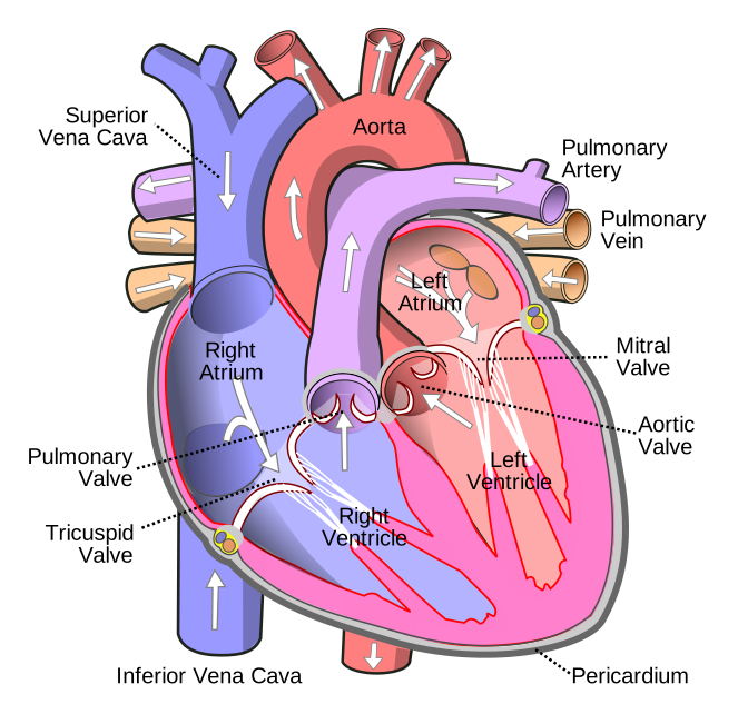
Two atria — thin-walled receiving chambers. Two ventricles — thick-walled pumping chambers.
The right side handles deoxygenated blood; the left side handles oxygenated blood. They are completely separated in the adult, and any hole between them is a defect (Chapter 4.5).
The path, in order:
Body → superior and inferior vena cava → right atrium → tricuspid valve → right ventricle → pulmonary valve → pulmonary artery → lungs → pulmonary veins → left atrium → mitral valve → left ventricle → aortic valve → aorta → body.
Two naming points that catch people out and matter for reading any report.
The pulmonary artery carries deoxygenated blood, and the pulmonary veins carry oxygenated blood. Arteries are defined by direction — away from the heart — not by oxygen content. This is the only place in the body where they diverge.
Both atria contract together, and both ventricles contract together. The heart is not left-then-right; it is atria-then-ventricles.
Why the left ventricle is thicker
The right ventricular wall is about 3 to 5 millimetres thick; the left is about 12 to 15 millimetres — roughly three times.
The reason is the pressure each has to generate, and the reason for that is the resistance of the circuit each supplies.
The right ventricle pumps into the pulmonary circulation, which is short, low-resistance, and highly distensible. It only has to generate about 25 mmHg systolic.
The left ventricle pumps into the systemic circulation — every organ, and up to a brain a considerable distance above it against gravity. It has to generate about 120 mmHg systolic, roughly five times as much.
And both eject the same volume every beat. They must, or blood would accumulate in one circuit. Over a day, an imbalance of even 1 percent would flood the lungs within hours. The matching is automatic, and Chapter 7.4 explains the mechanism.
The left ventricle is also more nearly circular in cross-section, while the right is crescent-shaped and wraps partly around it. A circular chamber generates pressure more efficiently; a crescent moves volume with less force.
And this explains right heart failure secondary to lung disease. If the lungs' vessels are damaged — by chronic lung disease, or by repeated clots — the pulmonary pressure rises, and the right ventricle, built for low pressure, cannot cope. It thickens, then dilates, then fails, producing swollen legs, an enlarged liver and distended neck veins. This is cor pulmonale, and it is a heart problem caused entirely by the lungs (Chapter 21.1).
The valves
Four valves, all passive — they open and close because of pressure differences, not because anything moves them.
Atrioventricular valves, between atrium and ventricle:
- Tricuspid on the right, with three cusps.
- Mitral (bicuspid) on the left, with two. Named for its resemblance to a bishop's mitre.
These have cords attaching their free edges to muscular pillars in the ventricle wall. The cords do not open or close the valve. They stop it inverting. When the ventricle contracts at high pressure, the cords hold the cusps down so they cannot flip back into the atrium — like the guy lines on an umbrella.
If a cord ruptures, as can happen after a heart attack or in infection, the valve suddenly leaks massively, and acute severe mitral regurgitation causes rapid pulmonary oedema and is a surgical emergency.
Semilunar valves, at the exits:
- Pulmonary valve, right ventricle to pulmonary artery.
- Aortic valve, left ventricle to aorta.
These have three pocket-like cusps and no cords. They close by blood filling the pockets from above when the ventricle relaxes.
The fibrous skeleton — a ring of dense connective tissue around the valves — anchors them and, importantly, electrically insulates the atria from the ventricles. This forces the electrical signal to pass through one specific point, which is the basis of the conduction system in Chapter 7.3.
Heart sounds
"Lub-dub" is the valves closing.
The first sound (lub) is the mitral and tricuspid valves closing as the ventricles start to contract. The second sound (dub) is the aortic and pulmonary valves closing as the ventricles relax.
The sounds are the abrupt deceleration of blood and the vibration of the valve and surrounding structures — not the leaflets slapping together.
A murmur is turbulent flow. Normal flow is smooth and silent. Turbulence occurs when blood passes through a narrowed valve, leaks backward through a valve that should be shut, or passes through an abnormal hole.
And the timing tells you which valve. A murmur between the first and second sounds is systolic — the ventricles are contracting, so it is either a narrowed exit valve or a leaking entry valve. Between the second and the next first sound is diastolic. Where it is loudest, and where it radiates to, identifies which of the four.
The stethoscope positions are not over the valves themselves but downstream in the direction the blood is travelling, which is why the aortic area is listened to at the right of the sternum rather than the left.
Not all murmurs are disease. Innocent flow murmurs are common in children, in pregnancy and in anaemia, simply because flow is faster.
The two valve diseases that matter most
Aortic stenosis — a narrowed aortic valve. Usually degenerative calcification in the over-70s, or earlier in people born with a two-cusp rather than three-cusp valve, which affects 1 to 2 percent of people.
The left ventricle must generate far higher pressure to force blood through, so it thickens. Eventually it cannot compensate.
The classic triad is angina, fainting on exertion, and breathlessness — and the prognosis attached to each is stark and worth knowing: once angina appears, average survival without treatment is around 5 years; once fainting appears, around 3 years; once heart failure appears, around 2 years. This is why symptomatic aortic stenosis is an indication for valve replacement rather than watchful waiting.
Valve replacement is now often done without opening the chest — TAVI, in which a replacement valve is delivered on a catheter through an artery in the groin and expanded inside the old valve. It began as an option for people too frail for surgery and has extended progressively to lower-risk patients.
Mitral regurgitation — a leaking mitral valve, so blood flows backward into the left atrium during systole. The atrium enlarges, which predisposes to atrial fibrillation (Chapter 7.3), and the lungs become congested.
Rheumatic heart disease remains a major cause of valve disease worldwide, though it has become rare in wealthy countries. The mechanism is a cross-reaction: antibodies raised against streptococcal throat infection attack heart valve tissue that happens to resemble the bacterium. It is entirely preventable by treating streptococcal sore throat with penicillin, which is why that unglamorous intervention matters so much in places where the disease is still common.
The heart wall
Three layers.
Endocardium — the smooth inner lining, continuous with the lining of the blood vessels. A smooth surface is essential, because any roughness triggers clotting.
Myocardium — the muscle (Chapter 4.2). Arranged in spiral bundles, so contraction wrings blood out with a twisting motion rather than a simple squeeze — which is more efficient than a purely concentric contraction would be.
Epicardium — the outer layer, which is also the inner serous layer of the pericardium.
Infective endocarditis is infection of the endocardium, usually on a valve. Bacteria in the bloodstream settle on a damaged or artificial valve and form vegetations — clumps of bacteria, platelets and fibrin.
Three things make it dangerous. Vegetations break off and travel, causing strokes and abscesses in distant organs. The valve is destroyed, causing acute heart failure. And the vegetation shields the bacteria from antibiotics and immune cells, which is why treatment requires 4 to 6 weeks of intravenous antibiotics rather than a short course.
It is why people with prosthetic valves or previous endocarditis are given antibiotics before dental procedures — dental work releases mouth bacteria into the blood. The indications for that prophylaxis have narrowed considerably as evidence accumulated that routine everyday bacteraemia from brushing teeth is a larger cumulative exposure than an occasional procedure, and good dental hygiene now matters more than the pre-procedure antibiotic.
What the next page fixes
The heart beats without any instruction from the brain — a heart removed from the body continues beating, and a transplanted heart with no nerve supply at all works. Chapter 7.3 covers where the beat comes from, how it spreads through the muscle in the right order, and how to read the electrical record of it on an ECG.