Appearance
1.6 — Energy: ATP and What Oxygen Is Actually For
Ask why you breathe and the usual answer is that oxygen burns your food. That is close enough to be memorable and wrong enough to make everything afterwards confusing, because it suggests oxygen is doing something to the glucose. It is not. Oxygen never touches the glucose.
What actually happens is that glucose is taken apart in stages, and at each stage electrons are stripped off it and handed to carrier molecules. Those electrons then run down a chain of proteins in your mitochondria, releasing energy as they fall, and that energy is used to build ATP. Oxygen sits at the very bottom of the chain and its only job is to catch the electrons at the end, combining with them and some hydrogen to make water. Nothing more.
That is why cyanide kills in minutes. It does not stop you breathing, it does not stop your blood carrying oxygen, and a cyanide victim's blood stays bright red because the oxygen is still there. Cyanide blocks the last protein in the chain, so the electrons have nowhere to go, the whole chain backs up like traffic behind a blocked junction, and ATP production stops throughout the body. The oxygen is present and useless.
This page follows one glucose molecule from your bloodstream to the ATP it becomes.
ATP: the cell's currency

Chapter 1.2 introduced ATP as a nucleotide, and here is why it matters. Three phosphate groups sit in a row, and every one of them carries negative charge. Like charges repel, so those three groups are being held next to each other against their strong preference to fly apart — think of three magnets forced together with the same poles facing.
Break the last one off, giving ADP (adenosine diphosphate) and a free phosphate, and three things happen at once. The repulsion is relieved. The free phosphate can spread its charge over its own oxygens in a way it could not while attached. And both products get surrounded by water molecules more comfortably than the original could. All three favour the split, and together they release energy.
The standard figure is 30.5 kilojoules per mole, but inside a real cell, where ATP is kept far more abundant than ADP, the usable release is nearer 50 to 60 kilojoules per mole. That is the packet size of biological energy: enough to do useful work, small enough that nothing is wasted on a job that needs less.
And the cell holds almost none of it. Your entire body contains roughly 250 grams of ATP at any moment — about five minutes of supply at rest, and a few seconds during hard exercise. It works because the same molecules are recycled constantly: ADP and phosphate are rejoined into ATP as fast as ATP is spent. Over a day the total throughput is roughly your own body weight. ATP is not a battery you charge up; it is cash moving through a till.
The overall reaction, and where the stages sit
The complete oxidation of glucose is written:
\mathrm{C_6H_{12}O_6} + 6\,\mathrm{O_2} \;\longrightarrow\; 6\,\mathrm{CO_2} + 6\,\mathrm{H_2O}
Read aloud: one glucose plus six oxygen molecules gives six carbon dioxide plus six water. The energy released is about 2,870 kilojoules per mole of glucose — the same total whether you burn it in a flame or metabolise it in a cell, because the start and end points are identical and energy released does not depend on the route (Volume IV, Chapter 3.4).
The difference is entirely in the route. A flame releases all 2,870 kilojoules at once as heat, which would cook a cell instantly. Your cells release it in about thirty small steps, capturing roughly 30 to 34 percent of it as ATP and letting the rest go as heat — which, incidentally, is where your body temperature comes from.
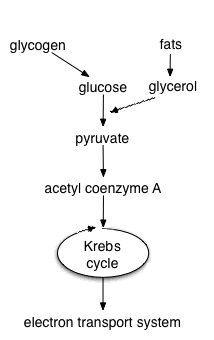
Four stages, in order:
- Glycolysis — in the cytosol, no oxygen needed, glucose split into two pyruvate.
- The link reaction — in the mitochondrial matrix, pyruvate converted to acetyl-CoA.
- The Krebs cycle — also in the matrix, acetyl-CoA fully taken apart into CO₂.
- Oxidative phosphorylation — in the inner mitochondrial membrane, where nearly all the ATP is actually made.
The electron carriers
Stages 1 to 3 barely make any ATP. What they make is loaded electron carriers, and you need two names.
NAD⁺ accepts two electrons and one proton to become NADH. It is built from niacin, vitamin B3. FAD accepts two electrons and two protons to become FADH₂. It is built from riboflavin, vitamin B2.
Think of them as rechargeable trays. Empty ones are loaded during the early stages, carried to the inner membrane, and emptied there — and the emptying is what makes the ATP. This is why a deficiency of B vitamins produces such devastating disease: without enough trays, the whole line stops regardless of how much food arrives.
Stage 1: glycolysis

Glyco — sugar. Lysis — splitting. Ten enzyme-catalysed steps in the cytosol, taking one six-carbon glucose to two three-carbon pyruvate molecules.
The first half spends energy. Two ATP are used to attach phosphate groups to the glucose. This looks backwards but does two necessary things: the added negative charge traps the molecule inside the cell, because a charged sugar cannot cross the membrane, and it destabilises the molecule so it is willing to be split.
The middle step splits it. The six-carbon molecule breaks into two three-carbon fragments, and everything after this point happens twice per glucose.
The second half collects. Each fragment is oxidised — electrons are stripped off onto NAD⁺, making NADH — and then rearranged so that phosphate groups can be handed directly to ADP. Two ATP per fragment, four in total.
Net result per glucose: 2 ATP, 2 NADH, 2 pyruvate. Spent two, made four, kept two.
Two features of glycolysis matter beyond the arithmetic.
It needs no oxygen and no mitochondria. This is why it is thought to be extremely old — every organism on Earth has it, including bacteria that have never seen oxygen, which suggests it was already in place before there was any oxygen to use. It is also why your red blood cells, which have no mitochondria at all, run entirely on glycolysis, and why a cell with its blood supply cut off can still make a little ATP for a while.
It is fast. Glycolysis produces ATP roughly a hundred times faster than the mitochondrial route, just far less of it per glucose. A muscle sprinting flat out uses this deliberately: low efficiency, high speed.
When oxygen runs short: fermentation
Glycolysis has a problem. It needs NAD⁺ to run, and it converts NAD⁺ to NADH. A cell holds only a small amount of NAD⁺, so unless NADH is emptied back to NAD⁺, glycolysis stops within seconds. Normally the mitochondria do the emptying. Without oxygen they cannot.
So the cell empties the trays itself. It takes the pyruvate and dumps the electrons from NADH straight onto it, converting pyruvate to lactate and regenerating NAD⁺. No further ATP is gained from this step. Its only purpose is to keep glycolysis running.
Yeast does the same trick with a different ending, converting pyruvate to ethanol and carbon dioxide. That reaction is bread rising, beer, and wine, and it is why all three are the same piece of biochemistry.
Two things people believe about lactate are wrong, and worth correcting because they change how you train.
Lactate does not cause the muscle soreness you feel two days after exercise. Blood lactate returns to normal within about an hour. The delayed soreness is microscopic damage to muscle fibres and the inflammation that follows it, which peaks at 24 to 72 hours. Chapter 24.5 covers it.
Lactate is not a waste product. It is a fuel. It leaves the muscle, travels in the blood, and is taken up by the heart, by other muscles and by the liver. The liver converts it back to glucose and returns it to the blood — a loop called the Cori cycle, which effectively lets the liver pay part of the energy debt of a sprinting muscle.
Where lactate does matter clinically is as a warning sign. A raised blood lactate in a sick patient means tissue somewhere is not getting enough oxygen, and it is one of the strongest predictors of death in sepsis. Chapter 17.11 uses it directly.
Stage 2: the link reaction
Pyruvate is transported into the mitochondrial matrix. There an enzyme complex called pyruvate dehydrogenase does three things to it in one operation: removes one carbon as CO₂, strips off electrons onto NAD⁺, and attaches the remaining two-carbon fragment to a carrier molecule called coenzyme A.
Per glucose (two pyruvates): 2 acetyl-CoA, 2 NADH, 2 CO₂.
That CO₂ is the first of the carbon dioxide you breathe out. It came from your food, not from the air.
This step is where thiamine, vitamin B1, is required as a coenzyme. Without it, pyruvate cannot enter the mitochondria's chemistry at all, and it piles up and is converted to lactate instead. The tissues that suffer first are the ones with the highest energy demand and the least ability to use fat — the brain and the heart. This produces beriberi: the "wet" form damages the heart and causes fluid overload, and the "dry" form damages nerves. In alcohol dependence, where thiamine intake is poor and absorption is impaired, the same deficiency produces Wernicke's encephalopathy, a medical emergency of confusion, unsteady walking and abnormal eye movements that can be reversed by intravenous thiamine and becomes permanent if it is not. This is why thiamine is given before glucose to a malnourished patient — giving glucose first raises demand for the enzyme and can precipitate the very crisis you were trying to treat. Chapter 24.2 gives the doses.
Stage 3: the Krebs cycle

Hans Krebs worked this out in 1937, and it is called a cycle because it ends where it began. The two-carbon acetyl group joins a four-carbon molecule called oxaloacetate to make six-carbon citrate — which is why it is also called the citric acid cycle. Then, around the ring, two carbons come off as CO₂ and electrons are stripped at four points, until the four-carbon oxaloacetate is regenerated and can accept the next acetyl group.
Per turn: 3 NADH, 1 FADH₂, 1 ATP (via GTP), 2 CO₂.Per glucose, which gives two turns: 6 NADH, 2 FADH₂, 2 ATP, 4 CO₂.
Add the earlier CO₂ and the books balance: 2 from the link reaction plus 4 here is 6, exactly the six carbons that walked in as glucose. The glucose is now completely dismantled. Every one of its carbons has left as carbon dioxide, and all its energy is sitting in loaded electron carriers.
The Krebs cycle also has a second job that gets less attention. It is a chemical crossroads. Fats enter it, protein fragments enter it, and molecules are drawn out of it to build amino acids and other things the cell needs. It is not just a furnace, it is the junction where all metabolism meets.
Stage 4: the electron transport chain and the machine that turns
Everything so far has produced 4 ATP per glucose. The remaining twenty-six or so come from here.

The chain. Four large protein complexes sit in the inner membrane. NADH delivers its electrons to Complex I; FADH₂ delivers to Complex II, which sits lower down and therefore yields less. Electrons pass from complex to complex, and at each transfer they drop to a lower energy level. Each complex has a stronger pull on electrons than the one before, so the electrons always move forward.
Where the released energy goes. Complexes I, III and IV use it to pump protons — hydrogen ions — from the matrix out into the space between the two mitochondrial membranes. The result is a proton concentration several times higher outside than inside, and, because protons carry charge, a voltage across the membrane as well. Peter Mitchell called this the proton motive force when he proposed the idea in 1961. It was ridiculed for a decade, because everyone was looking for a chemical intermediate rather than a gradient, and it won him the Nobel Prize in Chemistry in 1978.
The end of the line. At Complex IV the electrons are handed to oxygen, which combines with protons to form water. That is the entire purpose of the oxygen you breathe. It is the terminal electron acceptor — the thing at the bottom that accepts the rubbish so the chain can keep moving. Oxygen is used because it has an unusually strong pull on electrons, which makes the whole chain's energy drop as large as possible.

And now the elegant part. The protons want to come back in — the gradient is pushing them and the voltage is pulling them — but the membrane will not let charged particles through. The only route back is through ATP synthase, and ATP synthase is a genuine rotary motor.
Protons flow through a channel in its base, and as they do they force a ring of protein subunits to rotate, exactly as water turns a turbine. That ring turns a central shaft that runs up into the head of the enzyme. The head has three ATP-making sites arranged around it, and the shaft is asymmetric, so as it turns it deforms each site in sequence: one site is open and accepts ADP and phosphate, the next is squeezed shut and forces them to bond, the third is opened and releases the finished ATP. One full rotation of 360 degrees makes three ATP.
The motor runs at over a hundred revolutions per second. Paul Boyer worked out the mechanism and John Walker solved the structure, sharing the 1997 Nobel Prize in Chemistry. You are, in a completely literal sense, powered by billions of nanoscopic rotating engines.
The final count
Roughly 10 protons are pumped per NADH and 6 per FADH₂, and roughly 4 protons are needed per ATP made and exported. So each NADH is worth about 2.5 ATP and each FADH₂ about 1.5 ATP.
| Stage | ATP direct | NADH | FADH₂ | ATP from carriers |
|---|---|---|---|---|
| Glycolysis | 2 | 2 | — | ~3–5 |
| Link reaction | — | 2 | — | 5 |
| Krebs cycle | 2 | 6 | 2 | 18 |
| Total | 4 | 10 | 2 | ~26–28 |
About 30 to 32 ATP per glucose. Older textbooks say 36 or 38; those used round numbers of 3 and 2 ATP per carrier and ignored the cost of transporting things across the mitochondrial membrane. The modern figures are the ones above, and they are estimates rather than constants, because the true yield varies with conditions.
Efficiency check: 32 ATP at roughly 50 kilojoules each is about 1,600 kilojoules captured out of 2,870 available, around 55 percent under cellular conditions, or about 30 percent measured against the textbook standard value. Either way this is far better than a car engine, which manages 25 to 30 percent.
Fats and proteins take the same road
Glucose is not your main fuel at rest. Fat is.
Fatty acids are broken down in the mitochondria by beta-oxidation, which chops two carbons off the end of the chain at a time, producing one acetyl-CoA, one NADH and one FADH₂ per chop. The acetyl-CoA then enters the Krebs cycle exactly as it would from glucose. A 16-carbon fatty acid yields around 106 ATP — compare 32 from glucose, and you can see why fat is the storage form (Chapter 1.2).
When fat is being burned faster than the Krebs cycle can take it, as in prolonged fasting or uncontrolled diabetes, acetyl-CoA accumulates and the liver converts it to ketone bodies, which are water-soluble and can travel in blood to fuel the brain — which cannot use fatty acids directly, because they do not cross into the brain well. Mild ketosis during fasting is normal and useful. Diabetic ketoacidosis is not: in type 1 diabetes with no insulin, fat breakdown runs unchecked, ketones accumulate to the point of making the blood acidic, and the person becomes dangerously ill with vomiting, deep sighing breathing and a sweet acetone smell on the breath. It is a medical emergency, and Chapter 18.7 covers the recognition and the treatment.
Amino acids have their nitrogen removed first, and the remaining carbon skeletons enter at various points — some as pyruvate, some as acetyl-CoA, some directly into the Krebs cycle. The nitrogen becomes ammonia, which is toxic, so the liver converts it to urea and the kidneys excrete it (Chapter 10.2). This is why a blood urea level rises with a high protein intake, with dehydration, and with gastrointestinal bleeding, in which the blood in the gut is digested as protein.
What breaks it, and what that tells you
Cyanide binds Complex IV and stops the chain dead. Death follows in minutes, and the victim's skin often stays pink because the blood is still fully oxygenated. Treatment is hydroxocobalamin, a form of vitamin B12 that binds cyanide directly to form harmless cyanocobalamin excreted in urine, turning it a striking red. Chapter 23.6.
Carbon monoxide attacks the same system from two directions: it binds haemoglobin about 200 times more tightly than oxygen, so less oxygen is delivered, and it also inhibits Complex IV. It is odourless, and the classic cherry-red skin colour is unreliable. Chapter 23.6.
Uncouplers are the strangest failure mode. An uncoupler is a molecule that carries protons back across the inner membrane without going through ATP synthase. The gradient collapses, so no ATP is made, but the chain runs faster than ever trying to rebuild it, and all the energy comes out as heat.
2,4-dinitrophenol does exactly this, and it was sold as a weight-loss drug in the 1930s. It worked spectacularly — people lost weight rapidly because their food was being converted directly to heat. It also killed people, by cooking them from the inside with body temperatures above 43 °C, and caused cataracts. It was banned in the United States in 1938. It is still sold illegally online and still kills people every year, and there is no antidote. Anything that promises to make your metabolism burn fuel without doing work is describing a fever.
Your own body uses a controlled uncoupler on purpose. Brown fat contains a protein called UCP1, or thermogenin, which does the same thing deliberately to produce heat instead of ATP. Newborn babies are rich in it, because they cannot shiver effectively and lose heat fast through a large surface area relative to their volume. Hibernating animals use it to rewarm. Adults retain small amounts, mostly around the neck and shoulders, and it becomes more active in cold weather.
Where this shows up in your own life
Why you breathe harder when you run. Not because you need more air in your lungs, but because your muscles are drawing more electrons through their chains and need more oxygen at the end of them — and because the extra CO₂ produced is what your brain actually monitors. Chapter 8.5 explains why CO₂, not oxygen, is the signal that drives breathing.
Why a blocked coronary artery kills heart muscle in twenty minutes. Heart muscle cells are packed with mitochondria and have almost no capacity for glycolysis alone. Cut the oxygen and ATP collapses within minutes; the sodium pump of Chapter 1.4 stops; cells swell; and irreversible death begins at around 20 to 30 minutes, spreading outward over the following hours. This is the entire reason the phrase "time is muscle" exists, and why the goal in a heart attack is to reopen the artery as fast as physically possible. Chapter 18.3.
Why cold, fever and shivering all cost calories. All three are heat production, and heat production is chemical energy not captured as ATP.
And what a calorie on a food label really is. It is the total energy released by burning that food completely — the 2,870 kilojoules figure, converted to the older unit. Your body captures about a third of it as ATP and releases the rest as the warmth you can feel on your own skin right now.
What the next page fixes
Everything so far describes a cell running. It does not describe a cell making another cell — which is how you grew from one cell to thirty trillion, how your gut lining replaces itself every few days, and how a wound closes. It is also, when its controls fail, how cancer starts. Chapter 1.7 covers the cell cycle, mitosis and meiosis, and the checkpoints that decide whether a cell is allowed to divide at all.