Appearance
8.3 — The Mechanics of Breathing
The lungs contain no muscle of their own and cannot inflate themselves. They are inflated by making the chest bigger and letting atmospheric pressure push air in. You never suck air into your lungs; you lower the pressure inside them and the air outside does the rest.
That is why a hole in the chest wall is so dangerous, why you can only hold your breath for so long, and why a person struggling to breathe uses their neck and shoulder muscles.

The pressures
Three pressures matter, and all are measured relative to atmospheric pressure, which is taken as zero.
Alveolar pressure — inside the alveoli. Zero at the end of a breath in or out, when no air is moving. It goes slightly negative during inspiration (about −1 cmH₂O) and slightly positive during expiration.
Intrapleural pressure — in the pleural space. Always negative in normal breathing: about −5 cmH₂O at rest, −8 at full inspiration.
Transpulmonary pressure — the difference between alveolar and intrapleural pressure. This is the pressure holding the lung open, and it must stay positive or the lung collapses.
One breath
Inspiration is active.
- The diaphragm contracts and flattens, increasing the vertical dimension. The external intercostals lift the ribs, increasing the front-to-back and side-to-side dimensions (Chapter 5.4).
- Chest volume increases, so intrapleural pressure becomes more negative — from −5 to about −8.
- Because the lung is coupled to the chest wall by the pleural surface tension (Chapter 8.2), the lung is pulled open with it.
- Alveolar volume increases, so alveolar pressure falls below atmospheric — about −1 cmH₂O.
- Air flows in, because gases move from high pressure to low.
- As the lung fills, alveolar pressure returns to zero and flow stops.
Expiration at rest is passive. The muscles relax, the stretched elastic tissue of the lungs and chest wall recoils, volume falls, alveolar pressure rises to about +1, and air flows out. No energy is spent.
Forced expiration is active, using the abdominal muscles to push the diaphragm up and the internal intercostals to pull the ribs down. This is what you use for coughing, blowing, shouting and exercise.
Quiet breathing uses only about 3 to 5 percent of your total energy expenditure, and most of that is spent stretching elastic tissue against surface tension — which is exactly what surfactant reduces.
Why a hole in the chest wall collapses a lung
The negative intrapleural pressure is what holds the lung expanded. It exists because the lung's elastic recoil pulls inward while the chest wall's elasticity pulls outward, creating suction between them.
Open the pleural space to the atmosphere and that suction disappears instantly. Intrapleural pressure becomes zero, transpulmonary pressure becomes zero, and the lung's own elastic recoil collapses it toward its hilum. Meanwhile the chest wall springs slightly outward, which is why one side of the chest can look larger.
And this reveals something about the resting state. At the end of a normal breath out, the lungs are being held larger than they want to be, and the chest wall is being held smaller than it wants to be. They are in a permanent tug of war, and the pleural suction is the rope.
An open ("sucking") chest wound is treated with a dressing sealed on three sides, so air can escape but not enter — preventing a tension pneumothorax from developing while allowing the trapped air out. Chapter 23.8.
Compliance and resistance
Two properties determine how hard breathing is.
Compliance is how easily the lung stretches — volume change per unit pressure change. High compliance means floppy and easy to inflate; low compliance means stiff.
Low compliance (stiff lungs) — pulmonary fibrosis, pulmonary oedema, respiratory distress syndrome from lack of surfactant. Breathing is hard work on inspiration, and people compensate by taking rapid shallow breaths, because deep breaths are disproportionately expensive.
High compliance — emphysema, where elastic tissue is destroyed. This sounds like it should be good and is not. The lungs inflate easily but do not recoil, so expiration becomes the problem: air is trapped, the chest becomes hyperinflated, and the person has to force air out actively.
Resistance is the opposition to airflow, and it is overwhelmingly determined by airway radius (Chapter 7.5). Doubling the resistance requires double the pressure for the same flow.
High resistance — asthma, COPD, anything narrowing airways. The problem is expiration, because airways narrow naturally during expiration as the surrounding lung pressure rises.
This produces the single most useful distinction in a breathless patient. Restrictive disease is hard to breathe in; obstructive disease is hard to breathe out. Everything else follows: the pattern of breathing, the shape of the spirometry curve, and the treatment.
Lung volumes

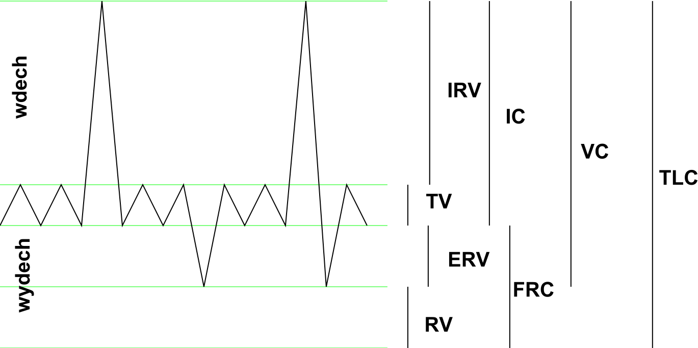
| Volume | Typical adult | What it is |
|---|---|---|
| Tidal volume | 500 ml | One quiet breath |
| Inspiratory reserve | 3,000 ml | Extra you can breathe in |
| Expiratory reserve | 1,100 ml | Extra you can breathe out |
| Residual volume | 1,200 ml | Cannot be expelled |
| Vital capacity | 4,600 ml | Maximum in to maximum out |
| Total lung capacity | 5,800 ml | Everything |
| Functional residual capacity | 2,300 ml | Left after a normal breath out |
Residual volume cannot be measured by breathing out into a machine, because by definition it cannot be exhaled. It needs helium dilution or body plethysmography.
And it is essential rather than wasted. It keeps the alveoli open between breaths, so they do not have to be reinflated from collapse every time. And it means gas exchange continues throughout the breathing cycle rather than in bursts — the functional residual capacity acts as a buffer that smooths the composition of alveolar gas, so arterial oxygen does not swing up and down with each breath.
Functional residual capacity is also why pre-oxygenation before anaesthesia works. Breathing 100 percent oxygen for three minutes replaces the nitrogen in that 2.3 litre reservoir with oxygen, giving several minutes of safe apnoea instead of under a minute. It is a standard step before every intubation and it is pure applied physiology.
Spirometry
Two measurements from one forced breath out:
FVC — forced vital capacity, the total volume exhaled. FEV₁ — the volume exhaled in the first second.
The ratio FEV₁/FVC is normally over 0.70 to 0.75.
Obstructive pattern — FEV₁ reduced much more than FVC, so the ratio falls below 0.70. Asthma, COPD, bronchiectasis. Air is in there; it just cannot get out fast.
Restrictive pattern — both reduced proportionally, so the ratio is normal or high. Fibrosis, obesity, chest wall deformity, muscle weakness. The lungs are small.
The reversibility test separates asthma from COPD. Measure, give a bronchodilator, measure again after 15 minutes. A significant improvement — usually more than 12 percent and 200 ml — indicates reversible obstruction, which is asthma. COPD is by definition largely irreversible, though many patients show some response.
Peak flow is a simpler measurement — the fastest rate achieved during a forced breath out. Its value is in tracking one person over time, which is why asthma action plans are built on personal best rather than population predicted values.
Work of breathing, and how to see it
Work of breathing is the energy spent moving air, and it is normally 3 to 5 percent of resting energy. In severe respiratory disease it can reach 30 percent or more, which is a genuinely important number: at that point the respiratory muscles are consuming a large share of the oxygen they are working to obtain, and the person is heading toward exhaustion.
The body minimises the work by choosing a breathing pattern, and the choice is informative.
Stiff lungs (restrictive) — inspiration is expensive, so rapid shallow breathing minimises the work. The cost is increased dead space ventilation (Chapter 8.1).
Obstructed airways — expiration is the problem, so slow deep breathing with a prolonged expiratory phase minimises the work.
And people with COPD often breathe out through pursed lips. This is not a habit; it is effective self-treatment. Pursed-lip breathing raises the pressure in the airways during expiration, splinting them open so they do not collapse and trap air. It genuinely improves emptying, and it is taught deliberately in pulmonary rehabilitation.
The signs of increased work of breathing are visible from across a room, and they are among the most useful observations in medicine because they need no equipment:
- Rapid breathing — over 20 breaths per minute in an adult.
- Use of accessory muscles — sternocleidomastoid and scalenes visible and active (Chapter 6.3).
- Intercostal and subcostal recession — the spaces between and below the ribs sucking in, especially visible in children.
- Nasal flaring in infants.
- Tripod position — sitting leaning forward on the arms, which fixes the shoulder girdle so the accessory muscles can pull on the ribs instead of the arms.
- Inability to speak in full sentences — one of the simplest and best severity markers in asthma.
And the most dangerous sign is the disappearance of these. A patient who was working hard and has become quiet, with a slowing respiratory rate and a silent chest, is not improving — they are exhausting. This is a well-recognised trap in severe asthma, and it is why "the silent chest" is taught as an ominous finding rather than a reassuring one.
Sighs, coughs and other manoeuvres
Sighing — a deep breath roughly every 5 to 10 minutes, largely unconscious. It reinflates alveoli that have gradually collapsed, and its absence is why patients on mechanical ventilation historically developed collapse until ventilators were programmed to deliver periodic larger breaths.
Coughing — a deep breath, closure of the glottis, forceful contraction of the expiratory muscles building pressure to around 100 mmHg, then sudden glottic opening. Air leaves at up to 800 kilometres per hour.
It requires all three components. A patient who cannot take a deep breath (pain, weakness), cannot close their glottis (a tracheostomy or an endotracheal tube), or cannot contract the abdominal muscles (pain, weakness, paralysis) cannot cough effectively — and cannot clear secretions. This is why pain relief after abdominal or chest surgery is a respiratory intervention (Chapters 5.4, 6.4).
Valsalva manoeuvre — forced expiration against a closed glottis. Used in lifting, defecation and straining. It raises intrathoracic pressure sharply, which reduces venous return — and that is why straining can cause fainting, and why it can be used deliberately to terminate a fast heart rhythm (Chapter 7.4).
Hiccup — Chapter 6.4.
Yawning — a deep breath with a wide mouth opening. Its function is genuinely unknown, and the popular explanation that it corrects low oxygen has been tested and does not hold: breathing high-oxygen or high-carbon-dioxide air does not change yawning rate. Brain cooling and a role in arousal or social signalling are the current candidates, and it is a fair example of a completely universal behaviour that science has not explained.
What the next page fixes
Air is now moving in and out. Chapter 8.4 covers what happens to it — how oxygen and carbon dioxide actually cross the barrier, how they are carried in blood, and why the oxygen dissociation curve's S-shape is one of the most elegant pieces of chemistry in the body.