Appearance
5.7 — The Lower Limb
The lower limb has the same plan as the upper — one bone, then two, then a cluster of small bones, then digits. Every part of it has been modified for load-bearing rather than manipulation, and the differences are instructive: the femur is enormously stronger than the humerus, the two leg bones cannot rotate over each other, and the foot's digits have lost almost all independent movement in exchange for forming an arch.
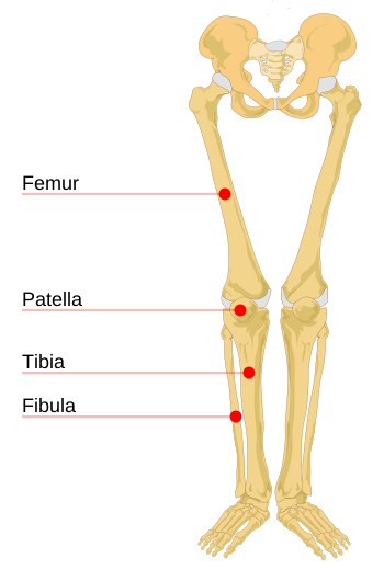
The femur
The longest, heaviest and strongest bone in the body, typically about 26 percent of a person's height — which is why it gives the best estimate of stature from skeletal remains (Chapter 5.1).
Its shaft can withstand compressive loads of well over 1,700 kilograms before failing, and during running the forces through it reach several times body weight.
It is not vertical. The femur angles inward from hip to knee, so that the knees sit under the body's centre line rather than under the hips. The angle is greater in females, because the acetabula are further apart (Chapter 5.6), and this has consequences at the knee.
The neck joins the head to the shaft at an angle of about 125 degrees. This angle is a lever arm, and it is the reason the hip abductor muscles have to work so hard. Standing on one leg, the body's weight acts at the midline while the supporting hip is off to the side, creating a turning force that would tip you over. The abductors on the standing side must pull down to balance it, and because their lever arm is short compared with the body weight's, the force through the hip joint when standing on one leg is around two and a half to three times body weight. Walking raises it further.
That is also why a person with weak or painful hip abductors lurches. They shift their whole trunk over the standing leg to move the body's centre of mass closer to the hip, reducing the turning force. The lurching gait is not weakness of the leg — it is a mechanical compensation, and it is a recognised clinical sign.
A fractured femoral shaft loses a lot of blood — 1 to 1.5 litres into the thigh — and it is painful enough to cause shock on its own. Traction splints are applied at the scene because pulling the leg straight reduces the volume the thigh can hold and takes the spasm out of the powerful thigh muscles, which reduces both bleeding and pain.
The patella
The kneecap is a sesamoid bone — one formed inside a tendon. It is the largest in the body, and it sits within the tendon of the quadriceps as it crosses the front of the knee.
Its function is purely mechanical: it increases the lever arm. By holding the tendon further forward from the joint's centre of rotation, it increases the turning force the quadriceps can produce by around 30 percent. Remove the patella and knee extension strength falls substantially, which is why patellectomy is now avoided wherever possible.
Its underside is covered with the thickest cartilage in the body, because the pressure between patella and femur during a deep squat reaches several times body weight.
Anterior knee pain — pain at the front of the knee, worse on stairs and after prolonged sitting — is extremely common, particularly in adolescents and young women, and is usually a problem of how the patella tracks in its groove rather than of any structural damage. It responds to strengthening the quadriceps and hip muscles far better than to anything else, and imaging usually shows nothing.
The tibia and fibula
The tibia is the shin bone and it carries essentially all the weight. It is the second longest bone in the body.
Its front border lies immediately under the skin for its entire length, with no muscle covering it. This is why a shin injury hurts so much and why open fractures of the tibia are common — there is nothing between the bone and the outside world. It is also why the tibia is a standard site for intraosseous access in emergencies (Chapter 5.1): the flat surface just below the knee is easy to find and to penetrate.
The fibula is slender and carries only about 10 to 15 percent of the load. Its main jobs are muscle attachment and forming the outer part of the ankle joint.
Because it carries so little weight, sections of fibula can be removed and used to reconstruct bone elsewhere — most commonly the jaw after cancer surgery, transplanted with its blood vessels and reconnected. The donor leg functions normally.
Unlike the forearm, the tibia and fibula do not rotate over each other. The bones are bound tightly together, and this is the trade: no pronation and supination in the leg, but a much more stable platform.
The common peroneal nerve wraps around the neck of the fibula, immediately under the skin, just below the outer side of the knee. This is the most commonly injured nerve in the leg, and the causes are ordinary: a plaster cast pressing there, sitting with legs crossed for a long time, prolonged squatting, or lying still during surgery. The result is foot drop — inability to lift the front of the foot, so the toes catch on the ground and the person has to lift the knee high to clear them. It is why casts and splints are padded specifically at that point.
The knee

The largest and most complex joint in the body, and structurally one of the most vulnerable, because it is essentially two rounded femoral condyles resting on a nearly flat tibial surface. The bone geometry provides almost no stability.
Four ligaments do the work.
Anterior cruciate ligament (ACL) — runs from the back of the femur to the front of the tibia, preventing the tibia sliding forward and controlling rotation. Posterior cruciate ligament (PCL) — the reverse, preventing backward slide. They cross inside the joint, which is what "cruciate" means. Medial collateral ligament (MCL) — resists force from the outside pushing the knee inward. Lateral collateral ligament (LCL) — resists the opposite.
Two menisci — C-shaped wedges of fibrocartilage that deepen the tibial surface, spread load over a larger area, and absorb shock.
The ACL is the most consequential ligament injury in sport. The mechanism is usually non-contact: landing from a jump or changing direction with the knee slightly bent and the foot planted. Around 70 percent of ACL tears involve no contact with another player at all. People typically report a pop, immediate swelling within a few hours from bleeding inside the joint, and a knee that feels unstable.
The ACL heals poorly because its blood supply is minimal and it sits in joint fluid that washes away any clot. Reconstruction uses a graft, usually from the person's own hamstring or patellar tendon.
Female athletes tear it two to eight times more often in the same sports, and the reasons are several: the greater angle from hip to knee (Chapter 5.6), differences in how the muscles activate on landing, a narrower notch in the femur through which the ligament runs, and hormonal effects on ligament laxity. Neuromuscular training programmes — teaching landing and cutting technique — reduce the injury rate by around half, which is one of the better-evidenced interventions in sports medicine.
Meniscal tears produce pain, swelling and sometimes locking, when a torn fragment jams in the joint. The outer third of a meniscus has a blood supply and can heal or be repaired; the inner two thirds do not and cannot. This is why some tears are repaired and others trimmed. And in older patients with degenerative tears, several large trials have found arthroscopic surgery no better than exercise therapy — a finding that has substantially reduced the number of these operations performed.
The "unhappy triad" — ACL, MCL and medial meniscus torn together — happens because a blow to the outside of the knee stresses all three at once.
The foot

26 bones: 7 tarsals, 5 metatarsals, 14 phalanges.
Tarsals: calcaneus (heel), talus, navicular, cuboid, and three cuneiforms.
The talus is unusual in two ways. No muscle attaches to it — it is moved entirely by the bones around it. And its blood supply enters from below and behind and is precarious, so a talar fracture frequently leads to avascular necrosis, exactly as with the scaphoid (Chapter 5.5).
The calcaneus is the largest tarsal and takes the impact of heel strike. A fall from height onto the heels is a characteristic injury pattern, and it should always prompt examination of the spine, because the force travels up the skeleton and lumbar compression fractures frequently accompany it.
The arches
The foot is not flat. It has a medial longitudinal arch (the high one on the inner side), a lateral longitudinal arch (low, on the outer side), and a transverse arch across the metatarsals.
The arches do three things. They act as a spring, storing energy on landing and returning it on push-off — around 17 percent of the energy of a running stride. They distribute load, so weight passes through the heel and the metatarsal heads rather than the whole sole. And they adapt: the foot flattens on contact to absorb shock and stiffens at push-off to become a rigid lever.
They are held by the shapes of the bones, by ligaments, and by the plantar fascia — a thick band running from the heel to the toes along the sole.
Plantar fasciitis is inflammation and degeneration where that band attaches to the heel. The characteristic complaint is sharp heel pain on the first steps in the morning, improving after walking and returning after rest — which happens because the fascia shortens overnight and is stretched abruptly on standing. It affects around 10 percent of people at some point. Treatment is calf and fascia stretching, supportive footwear, and time, and it resolves in the great majority within a year, though a year is a long time to be told to wait.
Flat feet are common and usually harmless if flexible — meaning an arch appears when standing on tiptoe. A rigid flat foot, where no arch appears, warrants investigation.
Achilles tendon — the strongest and thickest tendon in the body, joining the calf muscles to the calcaneus. It withstands loads of up to about 12 times body weight during running. Rupture typically happens in people in their thirties to fifties doing intermittent sport, and the description is consistent enough to be diagnostic: a sudden sensation of being kicked or struck in the back of the ankle, an audible snap, and inability to push off. The test is to squeeze the calf with the person lying face down — normally the foot points downward; with a complete rupture it does not move.
And fluoroquinolone antibiotics such as ciprofloxacin carry a genuine risk of tendon rupture, particularly the Achilles, particularly in the elderly and those on steroids. It is one of the reasons these antibiotics are now reserved rather than used routinely (Chapter 22.6).
Walking, in one paragraph
The gait cycle from one heel strike to the next has a stance phase (about 60 percent, foot on the ground) and a swing phase (about 40 percent). Both feet are on the ground during two brief overlaps — and running is defined precisely by the loss of those overlaps, replaced by a flight phase where neither foot is down.
Walking is remarkably efficient, because the body's centre of mass rises and falls like an inverted pendulum, exchanging potential and kinetic energy so that much of the energy is recovered rather than spent. Running is not a pendulum but a spring, storing energy in the Achilles tendon and the plantar fascia and returning it.
Any painful condition of the leg produces an antalgic gait — shortening the time spent on the painful side. It is one of the first things a clinician notices, before any examination, and it localises the problem to a side before a word is spoken.
What the next page fixes
Bones do nothing on their own. They are levers, and what moves them is the joints between them and the muscles crossing those joints. Chapter 5.8 covers joints — the three types, the six shapes of the movable ones, what cartilage is and why it does not heal, and what synovial fluid actually does.