Appearance
2.4 — Gene Regulation and Epigenetics
A liver cell and a neuron in your body contain identical DNA. The liver cell makes albumin, clotting factors and the drug-processing enzymes of Chapter 1.5. The neuron makes ion channels and neurotransmitter machinery, and never makes a single molecule of albumin — despite carrying a perfectly good albumin gene.
Roughly half of your 20,000 genes are switched on in any given cell type. The difference between your cells is not which genes they have. It is which genes they read. That control system is what this page is about, and it is the reason a cell can remain a liver cell through thousands of divisions without ever being told again.
Regulation in bacteria: the switch that started it
The first gene switch to be understood was in E. coli, and it is worth going through because it introduces every idea in miniature.
E. coli prefers glucose. If lactose is available and glucose is not, it needs three proteins: one to bring lactose into the cell, one to split it, and one accessory enzyme. Making them all the time would be wasteful, since lactose is usually absent.
The three genes sit together in a row with a single shared control region — an arrangement called the lac operon, described by François Jacob and Jacques Monod in 1961, for which they received the Nobel Prize in 1965.
When lactose is absent, a repressor protein sits on the DNA just downstream of the promoter, physically blocking RNA polymerase. Nothing is transcribed.
When lactose appears, a modified form of it binds the repressor and changes its shape so it can no longer grip the DNA. The repressor falls off, polymerase runs, and the three enzymes are made within minutes.
Two general principles come out of this.
First, the default state matters. The lac operon is off by default and switched on by need — a negative control. Other genes are on by default and switched off. Whether a system is normally on or normally off tells you what it costs to run and how urgent it is.
Second, the signal is a shape change in a protein. A small molecule binds a regulatory protein and alters its shape, and the shape decides whether it grips DNA. This is the same logic as the receptors in Chapter 1.8, and it is how nearly all regulation works.
Regulation in you: several layers, all at once
Human regulation is far more elaborate, because a human cell has to make a decision that lasts a lifetime rather than a decision that lasts twenty minutes. Control happens at every stage from DNA to finished protein.
1. Is the DNA even reachable?
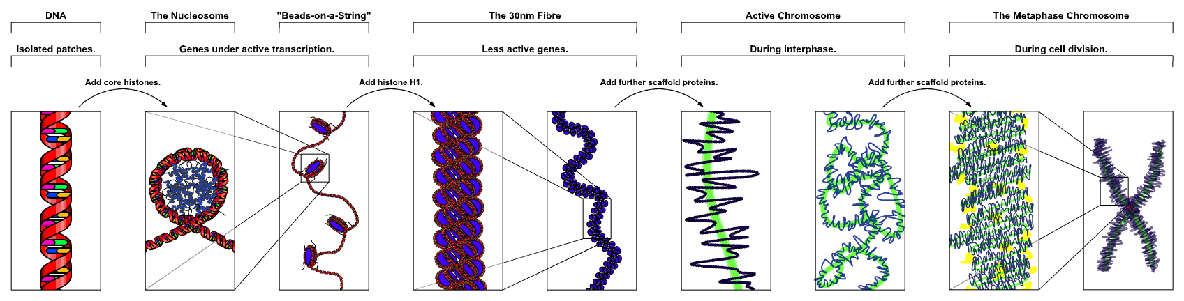
Two metres of DNA in a six-micrometre nucleus is not stored loose. It is wound around protein spools called histones, about 147 base pairs per spool, making a repeating unit called the nucleosome — the beads-on-a-string in the figure. Those are then coiled and looped further.
The tightness of that packing is the first and coarsest gene switch. DNA wound tight cannot be reached by RNA polymerase, so a gene in tightly packed chromatin — called heterochromatin — is off regardless of anything else. Loose chromatin — euchromatin — is available.
And the tightness is adjustable. Histones have flexible tails sticking out, and enzymes attach chemical groups to those tails.
Acetylation loosens. An acetyl group neutralises a positive charge on the histone, so its grip on the negatively charged DNA weakens and the DNA loosens. Acetylation is generally an "on" mark.
Methylation of histones can go either way, depending on exactly which amino acid is marked — some methyl marks close chromatin, others open it.
These marks are added by "writer" enzymes, removed by "erasers", and interpreted by "readers". The whole system is a drug target: histone deacetylase inhibitors such as vorinostat are used in certain lymphomas, and they work by preventing the removal of the loosening mark, forcing genes back on that a cancer cell had silenced.
2. Which transcription factors are present?
A transcription factor is a protein that binds a specific DNA sequence and either recruits RNA polymerase or blocks it. Humans have roughly 1,600 of them. Each gene's control region carries binding sites for several, so a gene is switched on only when the right combination is present — which is how a modest number of factors can specify a very large number of distinct cell types.
Control regions come in two kinds. The promoter sits immediately upstream of the gene. Enhancers can sit thousands or even a million bases away, and work because the DNA loops around to bring them next to the promoter. This is why a mutation far outside a gene can still break it: it can destroy an enhancer.
A dramatic demonstration: a single transcription factor called MyoD, forced into an ordinary skin fibroblast in culture, converts it into a muscle cell. One protein flips a master switch and a whole programme follows. This is the principle Yamanaka used with four factors to make induced pluripotent stem cells (Chapter 1.7).
3. What happens to the message after it is made?
- Alternative splicing decides which protein version is produced (Chapter 2.3).
- Message stability is set by the poly-A tail and by sequences in the untranslated regions.
- MicroRNAs are short RNA molecules that pair with a matching sequence in a message and either block its translation or trigger its destruction. Humans make well over a thousand of them, and a single microRNA can regulate hundreds of genes at once.
4. What happens to the protein after it is made? Phosphorylation switches enzymes on or off in seconds. Ubiquitin tagging sends them to the proteasome (Chapter 1.5). Cutting converts an inactive precursor into an active protein, as with insulin.
The layers differ in speed, and that is the point. Phosphorylating a protein takes seconds. Making new mRNA takes minutes. Repacking chromatin takes hours to days. A cell has fast switches for moment-to-moment adjustment and slow switches for permanent decisions, and the slow ones are what identity is made of.
Epigenetics: marks that outlast the signal
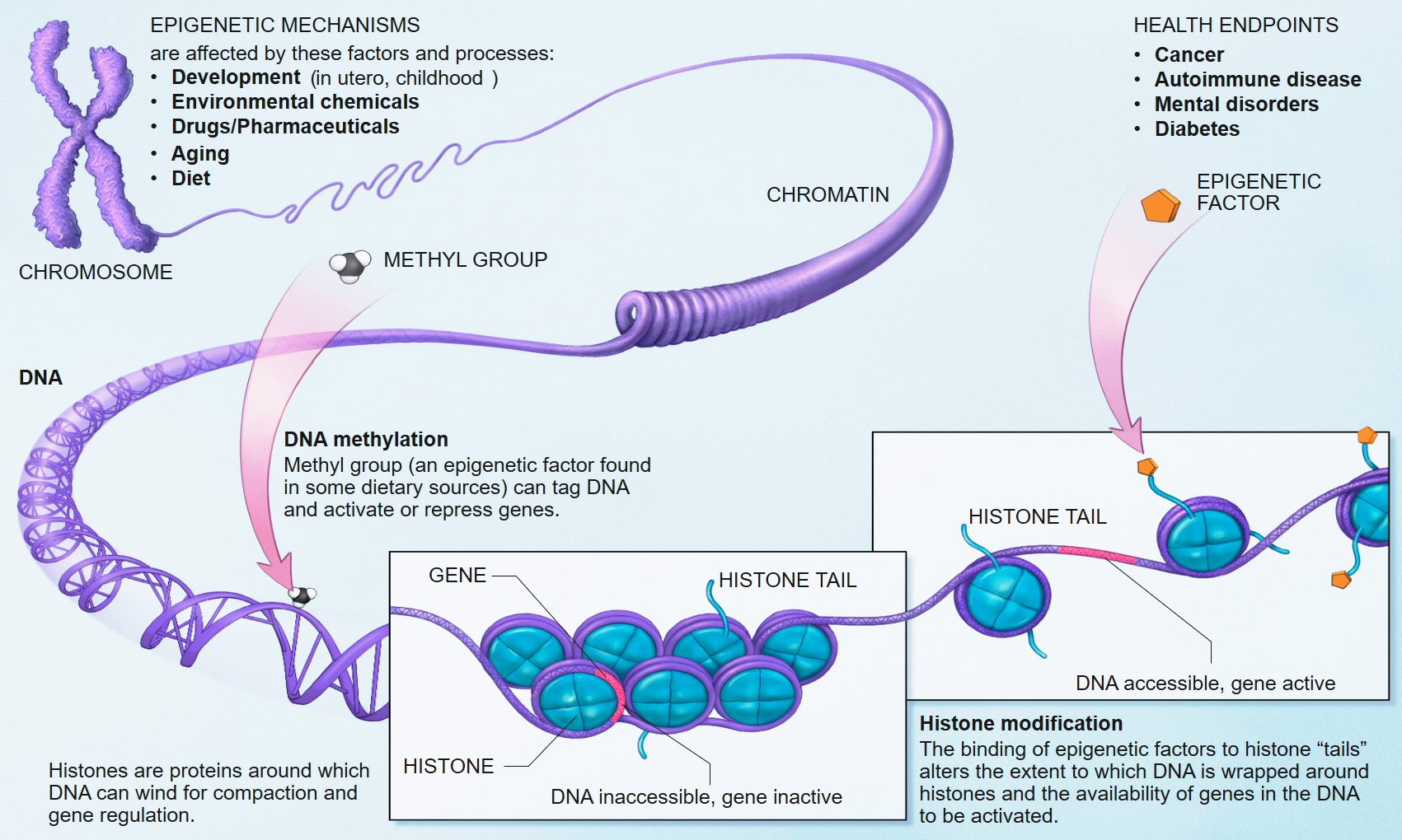
Epigenetics means, literally, "on top of genetics" — heritable changes in gene activity that do not change the DNA sequence. Two mechanisms carry most of the weight.
DNA methylation. A methyl group is attached directly to cytosine, almost always where a C is followed by a G in the sequence. Regions rich in this pattern, called CpG islands, sit at the front of many genes. A heavily methylated promoter is a silenced gene — the methyl groups recruit proteins that pack the chromatin shut.
And methylation is copied through cell division. When DNA is replicated, the new strand starts unmethylated, but an enzyme recognises the half-methylated pattern and completes it on the new strand. So the memory of "this gene is off" survives division without any signal being repeated. That is how a liver cell's daughters are liver cells.
Histone modification, described above, is the other mechanism, and it is partly copied through division too.
Three demonstrations that this is real and not hand-waving
X-chromosome inactivation. Females have two X chromosomes, males one. If both female Xs were fully active, the dose of every X gene would be double — which is not survivable. So early in development, each cell silences one X at random, packing it into a dense body called a Barr body, and every descendant of that cell keeps the same X silenced.
A tortoiseshell cat is this made visible. The gene for orange versus black coat colour sits on the X. A female cat carrying orange on one X and black on the other has patches: cells descended from those that silenced the black X are orange, and the reverse are black. The patch pattern is a map of which X was switched off in which founding cell. Male cats have only one X, which is why tortoiseshell males are essentially nonexistent — and the rare ones are XXY, the feline version of Klinefelter syndrome.
Genomic imprinting. For about a hundred human genes, it matters which parent a copy came from, because one parental copy is silenced by methylation laid down during egg or sperm formation. The clearest case is a region on chromosome 15. Lose the paternal copy of that region and you get Prader–Willi syndrome — low muscle tone in infancy, then insatiable appetite and obesity, short stature, and learning difficulties. Lose the maternal copy of the same region and you get Angelman syndrome — severe intellectual disability, absent speech, jerky movements and a characteristic happy demeanour. Same piece of DNA missing; two completely different diseases, decided only by which parent supplied the missing copy. Nothing in classical genetics predicts that.
The Dutch Hunger Winter. During the German blockade of the western Netherlands in the winter of 1944–45, the ration fell to around 400 to 800 calories a day. The Dutch kept meticulous medical records, so the children conceived during that famine have been followed for eighty years. Those exposed in early gestation had higher rates of obesity, diabetes, and cardiovascular disease as adults, and studies decades later found altered methylation at specific genes, including one involved in growth regulation. Some effects appear to extend to their children.
Be careful with how far that is pushed. The Dutch Hunger Winter findings are solid, but popular writing frequently extends epigenetics into claims about inheriting trauma across generations that the evidence does not currently support in humans. Most epigenetic marks are actively erased and rewritten in the early embryo — that reset is why a fertilised egg can become any cell type. Some marks at imprinted regions escape the reset; whether many others do in humans is genuinely unsettled.
Where this shows up in a hospital
Cancer is as much an epigenetic disease as a genetic one. Tumours very often silence tumour suppressor genes by methylating their promoters rather than mutating them — the gene is intact but switched off. This is important because it is potentially reversible. Azacitidine and decitabine are drugs that block DNA methylation, reawakening silenced genes, and they are used in myelodysplastic syndrome and some leukaemias. Chapter 19.7.
Methylation patterns are now a diagnostic tool. A tumour's methylation profile can identify which tissue it came from when the origin is unknown, and in brain tumours it has become a standard part of classification. In glioblastoma, whether a particular DNA repair gene's promoter is methylated predicts whether the drug temozolomide will work — because if the repair gene is silenced, the tumour cannot undo the damage the drug does.
And "epigenetic age" clocks measure methylation at a few hundred sites and estimate biological age with surprising accuracy. They predict mortality better than chronological age does. Whether they measure a cause of ageing or a consequence of it is not settled, and the commercial tests sold on this basis run far ahead of what is known. Chapter 24.8.
What the next page fixes
Regulation assumes the underlying sequence is intact. It is not always. Chapter 2.5 covers mutation — the kinds, what causes them, how the cell repairs them, and why a single deleted letter is usually catastrophic while a single substituted letter is usually harmless.