Appearance
10.2 — The Nephron and How Urine Is Made
The kidney's approach to excretion looks wasteful at first glance. It filters almost everything out of the blood indiscriminately, then carefully takes back what it wants. Filtering 180 litres a day to excrete 1.5 seems like an odd way to run a system.
It is actually the better design, and the reason is worth stating: filtering everything small and reclaiming the useful part means the kidney does not need a specific removal mechanism for every possible waste product, toxin or drug — including ones it has never encountered. Anything foreign that gets into your blood is filtered by default and excreted unless something actively reclaims it. A selective excretion system would have to be told about every new substance; this one handles them automatically.
The three processes
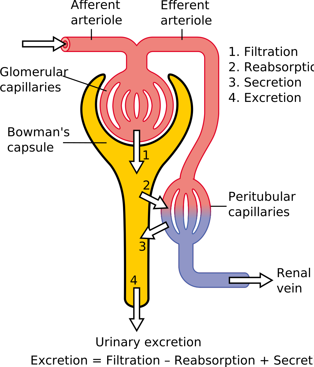
Filtration at the glomerulus — 180 litres a day into the tubule. Reabsorption from the tubule back to blood — about 178.5 litres. Secretion from blood into the tubule — for things that need removing faster than filtration alone achieves.
\text{Excreted} = \text{Filtered} - \text{Reabsorbed} + \text{Secreted}

Filtration
Glomerular filtration rate (GFR) is normally about 125 ml/min, which is 180 litres a day.
The driving pressure is the balance of Starling forces (Chapter 7.5):
\text{Net filtration pressure} = 55 - 15 - 30 = 10\ \text{mmHg}
Glomerular capillary pressure of 55 pushing out, capsular pressure of 15 pushing back, and plasma oncotic pressure of 30 pulling in.
Only 10 mmHg net — a small number producing an enormous flow, because the filter's surface area is large and its permeability is high.
Autoregulation keeps GFR nearly constant across mean arterial pressures from about 80 to 180 mmHg. Two mechanisms: the afferent arteriole constricts automatically when stretched by higher pressure, and the macula densa feedback from Chapter 10.1.
This is why moderate changes in blood pressure do not change urine output much, and why the kidney's function only falls when pressure drops below the autoregulatory range — which is exactly what happens in shock.
Measuring GFR is done indirectly, using creatinine — a waste product of muscle metabolism, produced at a fairly steady rate, freely filtered and only slightly secreted.
And its relationship with GFR is not linear, which is the single most misunderstood point in kidney medicine.
Because creatinine excretion equals production at steady state, the blood level is roughly inversely proportional to GFR. So:
| GFR | Approximate creatinine |
|---|---|
| 120 ml/min | 70 µmol/L |
| 60 ml/min | 140 µmol/L |
| 30 ml/min | 280 µmol/L |
Halving kidney function only doubles the creatinine, and the first doubling stays within or near the "normal" range. So a creatinine of 110 in a young woman may represent a 40 percent loss of function while still being reported as normal.
This is why estimated GFR is reported alongside creatinine, calculated from creatinine, age and sex. It is an estimate, and it is unreliable at the extremes of muscle mass — a bodybuilder's creatinine is high with normal kidneys, and a frail elderly person with little muscle can have a normal creatinine with badly impaired kidneys.
The proximal tubule: the bulk reclaim
About 65 to 70 percent of everything filtered is reabsorbed here, and most of it is not regulated — it simply happens.
| Substance | Reabsorbed here |
|---|---|
| Sodium | 65% |
| Water | 65% |
| Glucose | 100% |
| Amino acids | 100% |
| Bicarbonate | 80–90% |
| Potassium | 65% |
Sodium is the engine. The sodium–potassium pump on the basal side of the cell (Chapter 1.4) pumps sodium out into the blood, keeping intracellular sodium low. Sodium then flows into the cell from the tubule down that gradient — and that flow is used to drag other things with it.
Glucose, amino acids, phosphate and several other substances are all reabsorbed by sodium co-transport, exactly as in the gut. The sodium pump pays for all of it.
Water follows osmotically, and chloride follows electrically. So the fluid leaving the proximal tubule is much reduced in volume but almost the same concentration as plasma.
The transport maximum, and why diabetes causes sweet urine
Carriers can be saturated (Chapter 1.3). Glucose is normally reabsorbed completely, but only up to a limit.
The renal threshold for glucose is a blood level of about 10 to 11 mmol/L. Below it, all filtered glucose is reclaimed and none appears in the urine. Above it, the carriers are saturated and the excess spills through.
This is why diabetes was diagnosed for centuries by the taste of the urine, and why the name means "sweet siphon".
And it explains the classic symptoms in sequence. Glucose in the tubule is an osmotically active solute that cannot be reabsorbed, so it holds water in the tubule — an osmotic diuresis. The person passes large volumes of urine (polyuria), becomes dehydrated, and drinks constantly (polydipsia). The two cardinal symptoms of untreated diabetes are a direct consequence of one carrier being saturated.
And SGLT2 inhibitors work by lowering that threshold deliberately (Chapter 1.4), blocking the transporter so glucose is lost in the urine at normal blood levels.
Secretion, and where drugs go
The proximal tubule secretes hydrogen ions, and — importantly — organic acids and bases, including a large number of drugs.
Penicillin is secreted so efficiently that it is cleared from the blood very fast. During the Second World War, when penicillin was in critically short supply, it was recovered from patients' urine and re-purified for reuse. Probenecid, a drug that competes for the same secretory carrier, was developed specifically to block penicillin secretion and make each dose last longer — and it is still occasionally used for that purpose.
The same carrier system handles methotrexate, diuretics, uric acid and many other drugs, and competition between them at that carrier is a real source of drug interactions.
The loop of Henle: making the gradient
This is the mechanism that lets you make concentrated urine, and it is the most ingenious piece of engineering in the kidney.
The two limbs have opposite permeabilities, and that is the entire trick.
The descending limb is permeable to water and impermeable to salt. As it dips into the increasingly salty medulla, water leaves osmotically. The fluid inside gets more concentrated.
The ascending limb is impermeable to water and actively pumps salt out. Salt leaves and water cannot follow. The fluid inside gets more dilute — and the medulla around it gets saltier.
Now see how those two combine. The salt pumped out of the ascending limb makes the medulla salty. That saltiness draws water out of the descending limb. That makes the fluid entering the ascending limb saltier, so more salt can be pumped out.
Each step is small — the pump can only create a gradient of about 200 mOsm at any one level — but the countercurrent arrangement multiplies it along the length of the loop. The result is a gradient from about 300 mOsm at the top of the medulla to about 1,200 mOsm at the tip.
This is a countercurrent multiplier, and it is the same principle used in heat exchangers and in the legs of arctic birds. A modest local effect, repeated in series along two adjacent flows moving in opposite directions, produces a large overall gradient.
Urea contributes about half the gradient, recycled between the collecting duct and the loop — which is why a person with very low protein intake concentrates urine less well.
And the blood supply to the medulla has to be arranged not to wash the gradient away. The vasa recta run in hairpin loops alongside, so blood descending picks up salt and blood ascending gives it back. It is a countercurrent exchanger, preserving the gradient while still delivering oxygen.
Loop diuretics — furosemide — block the salt pump in the ascending limb. Two things follow. Salt is not reabsorbed, so it is lost in the urine with water following. And the medullary gradient collapses, so the collecting duct can no longer concentrate. This is why loop diuretics are the most powerful class, capable of producing very large urine volumes — and why they also cause loss of potassium, calcium and magnesium, which are all reabsorbed alongside in that segment.
The distal tubule and collecting duct: the fine adjustment
Only about 5 to 10 percent of the filtered load reaches here, and this is where all the regulation happens.
Aldosterone — from the adrenal cortex, in response to angiotensin II or high potassium. Increases sodium reabsorption and potassium secretion.
The two are linked, which explains a great deal of clinical medicine. Sodium is reabsorbed and potassium is pushed out in its place. So anything that raises aldosterone lowers potassium, and anything that blocks aldosterone raises it — which is exactly why spironolactone causes hyperkalaemia (Chapter 7.6) and why thiazide and loop diuretics cause hypokalaemia.
Antidiuretic hormone (ADH, vasopressin) — from the posterior pituitary, in response to concentrated blood or low volume.
Its mechanism is elegant: it inserts aquaporin water channels into the collecting duct membrane (Chapter 1.4). Without ADH the collecting duct is essentially waterproof; with it, water leaves osmotically into the salty medulla.
So the loop builds the gradient and ADH decides whether to use it.
Maximum concentration is about 1,200 mOsm/kg — four times plasma — allowing you to excrete the day's waste in as little as 500 ml. Minimum is about 50 mOsm/kg, allowing up to 20 litres a day if you drink that much.
Diabetes insipidus is the failure of this system, and there are two kinds.
Central — the pituitary does not make ADH. Nephrogenic — the kidney does not respond to it.
Either way the person passes enormous volumes of dilute urine — up to 20 litres a day — and is constantly thirsty.
The distinction between the two is made by giving synthetic ADH: the central form responds, the nephrogenic form does not.
And lithium is the commonest drug cause of the nephrogenic form, which is one of the reasons people on lithium need regular monitoring.
The name shares "diabetes" with diabetes mellitus because both cause enormous urine output; diabetes means siphon. One tastes sweet (mellitus, honeyed) and one does not (insipidus, tasteless) — a distinction made, before laboratories existed, exactly as it sounds.
What ends up in urine
Normal output: 1 to 2 litres a day.
Urea — from protein breakdown, and the main nitrogen waste. The liver makes it; the kidney excretes it (Chapter 9.4). Blood urea rises with a high protein intake, with dehydration, and with gastrointestinal bleeding, since blood in the gut is digested as protein.
Creatinine — as above.
Uric acid — from nucleic acid breakdown. Its accumulation causes gout (Chapter 21.5), and it is why gout is worsened by dehydration and by diuretics.
Electrolytes, hydrogen ions, ammonium, and various drugs and metabolites.
Colour tells you things. Pale means dilute. Dark yellow means concentrated — usually dehydration. Red or brown means blood or myoglobin (Chapter 6.7). Some drugs and foods colour it — beetroot, rifampicin (orange), and several others.
Foaming urine suggests protein. Cloudy urine suggests infection or crystals. A sweet smell suggests glucose; a strongly ammoniacal smell suggests infection.
Kidney stones
Affect around 10 percent of people at some point, more men than women, and recurrence without prevention is about 50 percent within ten years.
Types: calcium oxalate (about 80 percent), calcium phosphate, uric acid, struvite (from infection), and cystine (a rare inherited transport defect).
Formation requires supersaturation. Which means the single most important preventive measure is dilution, and it is remarkably effective: drinking enough to produce 2 to 2.5 litres of urine a day roughly halves recurrence. That one intervention outperforms most drugs.
Renal colic is the pain of a stone passing down the ureter — severe, coming in waves, radiating from loin to groin, with the person unable to keep still. It is frequently described as among the worst pains in medicine, and childbearing women who have had both often rate it comparably.
NSAIDs are more effective than opioids for renal colic, which is unusual and worth knowing. They reduce ureteric spasm and reduce the pressure in the obstructed system directly, rather than only blunting the perception.
Most stones under 5 millimetres pass spontaneously. Above 10 millimetres, most do not and need intervention — shock wave lithotripsy, ureteroscopy, or keyhole surgery.
A stone with fever is an emergency. An obstructed, infected kidney is effectively an abscess under pressure, and it can progress to sepsis within hours. It needs urgent drainage, not just antibiotics.
Prevention beyond fluid: reduce salt (which increases calcium loss into the urine), moderate animal protein, and maintain normal dietary calcium — this last point is counter-intuitive and important. Restricting dietary calcium increases stone risk, because calcium in the gut binds oxalate and prevents its absorption. Less dietary calcium means more oxalate absorbed and more excreted.
What the next page fixes
The nephron decides what stays and what goes. Chapter 10.3 covers what that decision is used for — holding your body water and every major electrolyte within ranges narrow enough that stepping outside them is a medical emergency, and what thirst actually is.