Appearance
7.8 — The Lymphatic System
About 20 litres of fluid leave your capillaries every day and only about 17 return directly (Chapter 7.5). The missing 3 litres would accumulate in your tissues within hours if nothing collected them, and complete blockage of lymphatic drainage is fatal within about a day.
The system that returns it is also the highway the immune system uses, the route by which fat is absorbed from your gut, and — unfortunately — the main path along which most cancers spread.
The structure
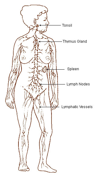
Lymphatic capillaries are blind-ended tubes in the tissues. Their structure is the elegant part. The endothelial cells overlap like roof tiles rather than forming tight junctions, and they are tethered to the surrounding tissue by fine filaments.
When tissue fluid accumulates, the swelling pulls on those filaments and opens the flaps between cells, letting fluid in. When pressure inside exceeds pressure outside, the flaps are pushed shut. So they are one-way valves made of cell edges, opening automatically in response to the very condition they exist to relieve.
And the gaps are large enough for proteins, cell debris, bacteria and whole cells to enter — which is exactly what makes lymph the immune system's collection route, and exactly what makes it the cancer spread route.
Lymphatic vessels are larger, with smooth muscle and one-way valves.
Lymph nodes — small bean-shaped organs, 1 to 25 millimetres, sited along the vessels. You have 500 to 600, concentrated at the neck, armpits, chest, abdomen and groin. Every lymph vessel passes through at least one node before returning to the blood.
The two ducts. Lymph eventually collects into two channels emptying into the large veins at the base of the neck.
The thoracic duct drains the entire lower body and the left side of the upper body — around three quarters of the body — into the left side. The right lymphatic duct drains only the right side of the head, neck, chest and right arm.
That asymmetry has a specific clinical use. Virchow's node is an enlarged lymph node above the left collarbone, and because the thoracic duct empties there, it can be the first sign of an abdominal cancer — most classically stomach cancer. A hard, fixed node in that position in an adult is a finding that triggers an urgent search for a primary tumour.
How lymph moves
There is no pump. The heart contributes nothing to lymph flow.
Four mechanisms move it, and all of them depend on movement:
Contraction of the vessels themselves. Lymphatic vessels have smooth muscle that contracts rhythmically, 6 to 10 times a minute, propelling lymph between valves.
Skeletal muscle pumping — the same mechanism as the venous pump (Chapter 6.6).
Breathing — pressure changes in the chest and abdomen with each breath.
Arterial pulsation — lymph vessels running alongside arteries are compressed with each pulse.
Total flow is only about 3 litres a day — roughly 2 ml per minute, against 5,000 ml per minute of blood. It is a slow, low-pressure system.
And because every mechanism depends on movement, immobility impairs lymphatic drainage, which is part of why immobile limbs swell and why elevation and movement are the first treatments for any swelling.
What lymph carries
Fluid and protein. Around 50 percent of plasma protein leaks into the tissues each day and must be returned. There is no other route back, which is why lymphatic obstruction causes such protein-rich, stubborn swelling.
Immune cells. Lymphocytes circulate continuously between blood, tissue, lymph and back — a cell may make this circuit once or twice a day, sampling for antigen as it goes (Chapter 13.2).
Antigens. Dendritic cells pick up material from tissues and travel through lymph to nodes, where they present it to T cells. This is where an immune response is initiated, which is why nodes swell during infection.
Fat. Fats absorbed from the small intestine do not enter the blood capillaries. They are packaged into chylomicrons — too large for blood capillaries — and enter specialised lymphatics in the villi called lacteals (Chapter 9.3).
This is why lymph from the gut is milky white after a fatty meal, which is where the word chyle comes from. And it is why damage to the thoracic duct — in chest surgery or trauma — causes chylothorax, milky fluid collecting in the chest, treated partly by putting the patient on a low-fat or fat-free diet so that less chyle is produced.
Lymph nodes
A node is a filter and a meeting place.
Lymph enters through several vessels, percolates through a network of channels lined with macrophages that remove bacteria, debris and cancer cells, and leaves through one vessel. Inside, B cells cluster in follicles and T cells occupy the surrounding zone.
Node enlargement — lymphadenopathy — is one of the commonest findings in clinical examination, and the character of the node tells you a great deal.
| Feature | Suggests reactive | Suggests malignant |
|---|---|---|
| Size | Under 1 cm | Over 2 cm |
| Consistency | Soft, rubbery | Hard, stony |
| Mobility | Freely mobile | Fixed to surroundings |
| Tenderness | Tender | Painless |
| Course | Resolves in weeks | Persists and grows |
A tender, mobile, soft node after a sore throat is reactive. A hard, painless, fixed, enlarging node in an adult is malignancy until proven otherwise, and the standard is that a node persisting beyond about six weeks without explanation is investigated.
Generalised lymphadenopathy — nodes enlarged in several regions — points to a systemic cause: infectious mononucleosis, HIV, tuberculosis, lymphoma, or an autoimmune disease.
Location narrows it. The node drains a specific territory, so an enlarged node points to its drainage area. Nodes above the collarbone are almost always significant. Nodes in the armpit drain the breast and arm. Nodes in the groin drain the leg and genitals — and are commonly enlarged for trivial reasons in people who walk barefoot or have minor foot injuries, which is worth knowing before alarming someone.
The spleen
The largest lymphoid organ, in the left upper abdomen behind ribs 9 to 11, weighing about 150 grams.
Two functionally distinct tissues:
Red pulp — filters blood. Macrophages remove aged and damaged red cells — this is where the 120-day lifespan ends (Chapter 7.1) — and recover the iron. It also stores platelets: around a third of your platelets are in the spleen at any moment.
White pulp — lymphoid tissue that mounts immune responses to blood-borne organisms.
The spleen's most important immune role is against encapsulated bacteria — organisms with a polysaccharide capsule that resists ordinary phagocytosis. The spleen is uniquely good at clearing them, and without it a person is dangerously vulnerable.
Splenic rupture is one of the commonest serious injuries in blunt abdominal trauma, because the spleen is fragile, highly vascular and only protected by the lower ribs. A fractured left lower rib should always prompt consideration of splenic injury. It bleeds heavily and rapidly, and can present with shoulder tip pain from blood irritating the diaphragm (Chapter 6.4).
Asplenia — living without a spleen — whether from surgical removal, trauma, or sickle cell disease destroying it (Chapter 2.8), carries a specific, lifelong and preventable risk.
Overwhelming post-splenectomy infection can progress from feeling unwell to death within 12 to 24 hours. The organisms are the encapsulated bacteria: pneumococcus, meningococcus, and Haemophilus influenzae type b.
The protective measures are simple and must be lifelong:
- Vaccination against all three organisms, plus annual influenza vaccination, ideally given at least two weeks before a planned splenectomy.
- Prophylactic antibiotics — usually penicillin, at least for the first few years and often lifelong.
- A supply of emergency antibiotics to start immediately at the first sign of fever, before seeing a doctor.
- A medical alert card or bracelet.
- Malaria precautions, because asplenic people get far more severe malaria.
This is one of the clearest cases in medicine where knowing one fact about yourself changes what you must do when you get a fever, and where the information is frequently not communicated adequately after the operation.
Lymphoedema
Swelling caused by lymphatic failure. It is distinct from other oedema in a way that is clinically recognisable: because protein cannot be cleared, the fluid is protein-rich, and over time it causes fibrosis and thickening of the skin.
So lymphoedema does not pit — pressing it does not leave an indentation — once it is established, whereas oedema from heart failure or low albumin does. And the skin becomes thickened and warty over years.
Primary lymphoedema — a developmental abnormality of the vessels, sometimes presenting at birth and sometimes in adolescence.
Secondary lymphoedema — far commoner.
Cancer treatment is the leading cause in wealthy countries. Removing axillary lymph nodes for breast cancer causes arm lymphoedema in a substantial proportion — historically 20 to 30 percent after full axillary clearance, and much less with modern techniques. Radiotherapy adds to it.
This risk is precisely why sentinel lymph node biopsy was developed. A dye or tracer is injected at the tumour and travels to the first node draining it — the sentinel node. If that node is clear, the rest almost certainly are too, and the remaining nodes are left in place. It has substantially reduced lymphoedema rates without compromising cancer outcomes, and it is one of the better examples of surgery becoming less invasive on the basis of anatomy.
Filariasis is the leading cause worldwide. Parasitic worms transmitted by mosquitoes lodge in the lymphatic vessels and obstruct them, causing massive swelling of the legs and genitals — elephantiasis. It affects tens of millions of people, and mass drug administration programmes have eliminated it from several countries.
Treatment of established lymphoedema is compression garments, manual lymphatic drainage massage, exercise, and meticulous skin care. The skin care matters because the limb is prone to cellulitis, and each episode of infection damages more lymphatics, making the swelling worse — a vicious circle that patients must be taught to interrupt by treating any break in the skin promptly.
There is no cure, which is why prevention through surgical technique matters so much.
Cancer spread
Most carcinomas spread through the lymphatics first (Chapter 19.3), and the reason is the anatomy at the top of this page: lymphatic capillaries have gaps large enough for a cell to enter, and no basement membrane to cross.
Cells travel to the first draining node, lodge there, and grow. From there they progress to the next nodes and eventually into the bloodstream.
This orderly progression is what makes lymph node status the single most important prognostic factor in most solid cancers, and it is why nodes are sampled during cancer surgery and why staging systems count them (Chapter 19.4).
Sarcomas behave differently — they spread through the blood rather than the lymphatics, which is why lymph nodes are not routinely removed in sarcoma surgery.
What the next page fixes
Everything in this Part has described the adult circulation. A fetus has no air to breathe and its lungs are collapsed and full of fluid, so sending blood to them would be pointless. Chapter 7.9 covers the fetal circulation — three shunts that bypass the lungs and liver, and the sequence of changes at the first breath that closes them all within minutes.