Appearance
10.1 — Kidney Anatomy
Your kidneys filter about 180 litres of fluid a day and give back 179 of them. The entire plasma volume of your body passes through them roughly sixty times a day. They weigh 150 grams each — under 0.5 percent of your body weight — and they take 20 to 25 percent of your cardiac output to do it.
They do not receive that blood because they need it. They receive it because processing the blood is the point.
Position and size

Retroperitoneal (Chapter 4.1), lying against the back wall between T12 and L3.
The right kidney sits about 1 to 2 centimetres lower than the left, because the liver occupies the space above it.
Each is about 11 to 12 cm long, 6 cm wide, 3 cm thick — roughly the size of a computer mouse, and about 150 grams.
Their position explains how they are examined and reached. They lie under the lower ribs at the back, so kidney pain is felt in the flank, in the angle between the last rib and the spine — the renal angle, and tenderness there is a specific sign of kidney inflammation. And a kidney biopsy is taken through the back, with the patient lying face down.
Three protective layers: a fibrous capsule directly on the kidney, a layer of perirenal fat, and an outer fascial envelope. The fat matters — it cushions the kidney and holds it in position, which is why very thin people can have a mobile kidney that drops when they stand.
The capsule is tough and does not stretch quickly. This is why anything that swells the kidney rapidly — acute inflammation, obstruction — causes pain, while slow enlargement does not.
Internal structure
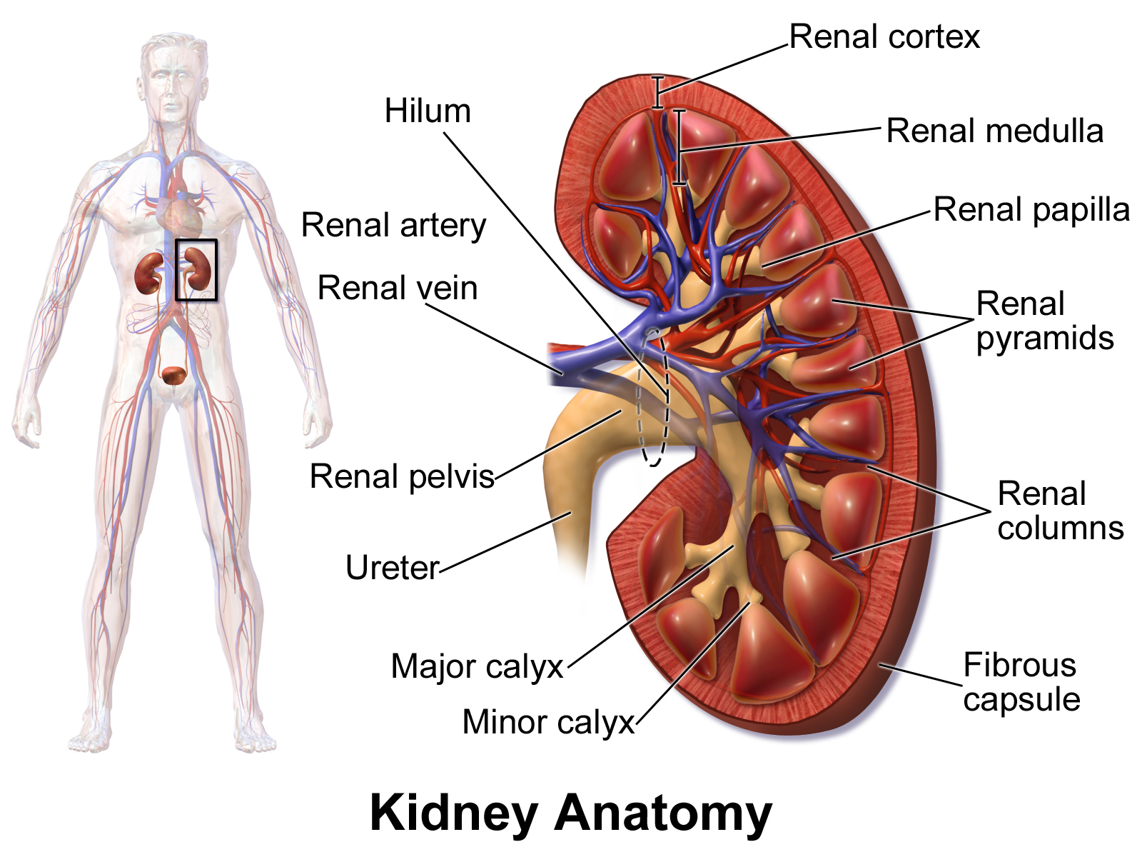
Cortex — the outer band, about 1 centimetre thick. Contains all the filtering units and most of the tubules.
Medulla — the inner region, arranged into 8 to 18 pyramids whose striped appearance comes from parallel tubules and blood vessels all running in the same direction. The tips point inward.
Calyces — cup-shaped collecting chambers around each pyramid tip. Minor calyces merge into major calyces.
Renal pelvis — the funnel where they all meet, narrowing into the ureter.
The hilum — the notch on the medial side where the artery enters, the vein leaves, and the ureter exits.
The order at the hilum is fixed and worth knowing: vein in front, artery in the middle, ureter behind. It matters in surgery, and it is the kind of small fact that stops a serious mistake.
The nephron
The functional unit — about one million per kidney.
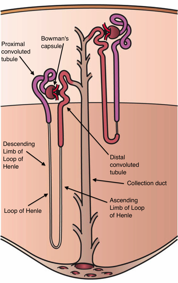
Glomerulus — a tuft of capillaries where filtration happens.
Bowman's capsule — the cup surrounding it, catching the filtrate.
Proximal convoluted tubule — where most reabsorption happens.
Loop of Henle — dips down into the medulla and comes back. This is what allows concentrated urine to be made, and Chapter 10.3 explains how.
Distal convoluted tubule — fine adjustment, under hormonal control.
Collecting duct — final water adjustment, then down through the medulla to the calyx.
Two kinds of nephron, and the proportion is significant.
Cortical nephrons (about 85 percent) have short loops staying mostly in the cortex.
Juxtamedullary nephrons (about 15 percent) sit at the cortex–medulla border with long loops plunging deep into the medulla. These are the ones that create the concentration gradient, and desert mammals have far more of them — a kangaroo rat can produce urine several times more concentrated than a human and never needs to drink.
You lose nephrons with age — roughly 10 percent per decade after 40, and they are not replaced. The reserve is so large that this rarely matters on its own, but it is why kidney function must be checked before prescribing certain drugs in older people, and why the same insult that a young kidney shrugs off can tip an older one into failure.
You can live normally with one kidney. The remaining one hypertrophies and takes over about 80 percent of the original combined function. This is why living kidney donation is possible, and donors have essentially normal life expectancy.
The blood supply, and the two capillary beds
This is the most unusual feature of renal anatomy, and everything about kidney function depends on it.
Renal artery → segmental → interlobar → arcuate → interlobular → afferent arteriole → GLOMERULUS → efferent arteriole → peritubular capillaries → veins.
Notice the anomaly. In every other organ, blood goes artery → capillary → vein. Here it goes artery → capillary → artery → capillary → vein. Two capillary beds in series, separated by an arteriole.
That arrangement is the whole trick, and it does two things.
The glomerular capillaries operate at high pressure, because they sit between two arterioles rather than draining into a vein. Glomerular capillary pressure is about 55 mmHg against 15 to 25 mmHg in an ordinary capillary. That high pressure is what drives filtration.
The peritubular capillaries operate at low pressure and high oncotic pressure, because plasma has just been concentrated by losing 20 percent of its water at the glomerulus. Low hydrostatic pressure plus high oncotic pressure is exactly the condition for reabsorption (Chapter 7.5). The same Starling forces that filter fluid out at the glomerulus draw it back in downstream.
One design, two opposite behaviours, achieved by putting an arteriole between them.
And the kidney can adjust filtration precisely by changing the two arterioles independently.
Constrict the afferent (incoming) — pressure in the glomerulus falls, filtration falls. Constrict the efferent (outgoing) — pressure in the glomerulus rises, filtration rises.
This is why ACE inhibitors and ARBs affect kidney function (Chapter 7.6). Angiotensin II preferentially constricts the efferent arteriole, maintaining filtration pressure. Block it and the efferent arteriole dilates, glomerular pressure falls, and the filtration rate drops.
Two consequences follow, and they pull in opposite directions. A small rise in creatinine — up to about 30 percent — when starting these drugs is expected and acceptable, and does not mean the drug is damaging the kidney. And in a patient with narrowing of both renal arteries, who depends entirely on that efferent constriction to filter at all, these drugs can precipitate acute kidney failure. Both facts follow from one arteriole.
Long term, the same effect is protective: reducing glomerular pressure reduces the damage that high pressure does to the filter over years, which is why these drugs slow the progression of diabetic and hypertensive kidney disease.
The medulla is relatively hypoxic, because the blood vessels running alongside the loops of Henle exchange oxygen with each other rather than delivering it deep. So the medulla operates on a thin oxygen margin, and it is the first part of the kidney to be damaged by low blood pressure — which is why shock causes acute tubular necrosis specifically.
The filter
Three layers between blood and filtrate, and each excludes something different.
Fenestrated endothelium — the capillary lining, riddled with pores about 70 to 100 nanometres across. Stops blood cells; passes everything smaller.
Basement membrane — a mesh of collagen and glycoproteins. The main size barrier, and it carries a strong negative charge.
Podocytes — remarkable cells with interdigitating foot processes wrapped around the capillary, leaving narrow slits between them bridged by a fine protein diaphragm. The final and most selective barrier.
Two properties select what passes.
Size. Molecules under about 10 kilodaltons pass freely. Above about 70 kilodaltons, essentially nothing passes.
Charge. The barrier is negatively charged, so negatively charged molecules are repelled. Albumin is 69 kDa — right at the size limit — and is also negatively charged, so it is doubly excluded. Less than 1 percent of filtered plasma protein appears in the filtrate.
So proteinuria — protein in the urine — means the filter is damaged, and it is one of the earliest and most useful markers of kidney disease.
In diabetes the charge barrier is lost before the size barrier, so small amounts of albumin appear first. This is why the earliest test for diabetic kidney disease is the urine albumin-to-creatinine ratio rather than a standard dipstick — the dipstick only detects larger losses, by which point damage is established. Detecting it at the microalbuminuria stage and starting an ACE inhibitor genuinely changes the long-term outcome, and it is one of the clearest cases where an early test alters a life.
Nephrotic syndrome is massive protein loss — over 3.5 grams a day — with low blood albumin, swelling and high cholesterol. The swelling is the oncotic pressure explanation from Chapter 7.1.
Nephritic syndrome is inflammation of the glomeruli, letting red cells through. Blood in the urine, some protein, high blood pressure and reduced filtration.
The juxtaglomerular apparatus
Where the distal tubule of a nephron loops back and touches its own afferent arteriole — a small structure with an outsized role.
Two cell types:
Juxtaglomerular cells in the arteriole wall, which sense pressure and secrete renin (Chapter 7.6).
Macula densa cells in the tubule wall, which sense the sodium chloride concentration of the fluid passing them.
Together they form a feedback loop that regulates a single nephron's own filtration rate. If filtration is too high, more sodium reaches the macula densa, which signals the afferent arteriole to constrict and reduce it. Each nephron regulates itself, a million times over.
And this same apparatus is the sensor for whole-body blood pressure control, because low pressure means low sodium delivery, which triggers renin release and the whole renin–angiotensin cascade.
The ureters, bladder and urethra
Ureters — about 25 to 30 centimetres, carrying urine by peristalsis. Gravity is not required, which is why you can lie down.
Three natural narrowings, and stones lodge at all three: where the ureter leaves the renal pelvis, where it crosses the pelvic brim over the iliac vessels, and where it enters the bladder wall.
The ureters enter the bladder obliquely, tunnelling through its wall. As the bladder fills, the rising pressure compresses that tunnel and seals it — a valve made of geometry rather than of muscle, preventing urine refluxing back toward the kidney. Failure of this arrangement causes vesicoureteric reflux in children, and repeated kidney infections and scarring.
Bladder — a muscular reservoir holding 400 to 600 ml comfortably. Chapter 10.5.
Urethra — about 4 centimetres in women and 20 centimetres in men.
That difference explains the epidemiology of urinary infection almost entirely. A short urethra with its opening close to the anus is why urinary tract infections are so much commoner in women — roughly 50 percent of women have at least one in their lifetime against a much smaller proportion of men. And a urinary infection in a man is treated as significant, because it usually implies an underlying abnormality.
What the kidney does besides making urine
Four jobs that have nothing to do with excretion, and their failure is why chronic kidney disease affects the whole body.
Erythropoietin — the hormone driving red cell production (Chapter 7.1). Its loss is why kidney failure causes anaemia, and synthetic EPO treats it.
Vitamin D activation — the final hydroxylation step happens in the kidney (Chapter 5.1). Its loss causes bone disease in kidney failure, and treatment requires the already-activated form.
Blood pressure control — renin, and long-term volume control (Chapter 7.6). Which is why kidney disease causes hypertension, and hypertension causes kidney disease — a loop that has to be broken pharmacologically.
Acid–base balance — Chapter 10.4.
What the next page fixes
The kidney filters 180 litres and returns 179. Chapter 10.2 follows the filtrate through the nephron segment by segment and shows exactly what is taken back where, why glucose appears in the urine only above a specific blood level, and how the loop of Henle achieves the concentration gradient that lets you produce urine four times more concentrated than your blood.