Appearance
11.5 — The Cerebrum, Lobe by Lobe
In 1848, a railway foreman named Phineas Gage survived a metre-long iron rod being blasted through his skull. It entered under his left cheekbone and exited through the top of his head, destroying much of his left frontal lobe. He was speaking within minutes and walked to the cart.
What changed was his personality. The reports — which have been embellished considerably over the years, and the reliable core of which is thinner than the popular version — describe a reliable, capable man becoming impulsive, profane and unable to hold to a plan.
The case established something genuinely new: that character and judgement have a physical seat, and that it could be damaged while leaving movement, speech and memory intact.
The cortex
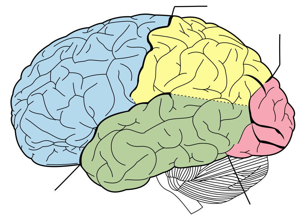
A sheet of grey matter 2 to 4 millimetres thick, folded so that about two thirds of it is buried in the grooves.
Unfolded it would cover about 2,500 square centimetres — roughly a large dinner napkin. The folding is what allows a large sheet in a small skull, and it is why species with more cortex have more folded brains.
The ridges are gyri and the grooves are sulci.
Six layers, each with characteristic cell types and connections. The layer structure differs by region in ways that correspond to function — which is how Brodmann divided the cortex into 52 numbered areas in 1909, purely from microscopic appearance, and those areas turned out to correspond remarkably well to functional divisions established decades later.
White matter beneath carries three kinds of fibre: association fibres connecting regions within one hemisphere, commissural fibres connecting the two hemispheres — of which the corpus callosum, with about 200 million axons, is by far the largest — and projection fibres running to and from the rest of the nervous system.
Deep grey structures sit within the white matter: the basal ganglia, which are central to initiating and selecting movement and are where Parkinson's disease and Huntington's disease act; and the limbic structures of Chapter 11.6.
The frontal lobe
The largest lobe, and the one that has expanded most in human evolution.
Primary motor cortex — the strip immediately in front of the central sulcus. Controls voluntary movement of the opposite side.
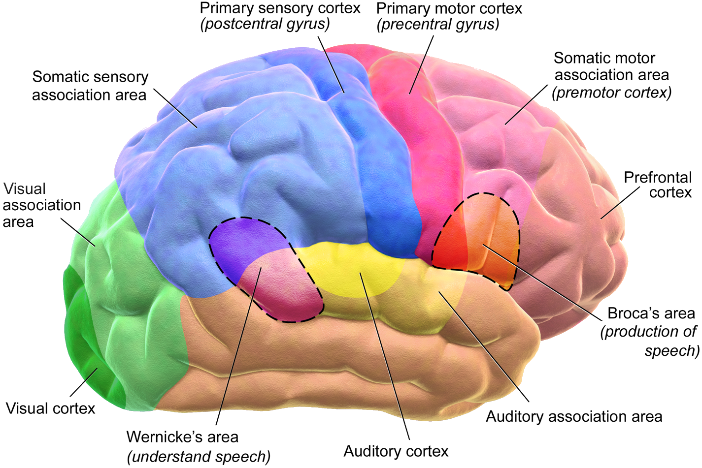
The homunculus — the body map — has three properties worth knowing.
It is inverted: the legs are at the top, near the midline, and the face at the bottom.
It is disproportionate: the hands and the lips and tongue occupy an enormous share, because they are the most finely controlled. The trunk and legs occupy very little.
And the leg area is on the medial surface, tucked into the midline between the hemispheres. This is why a stroke in the anterior cerebral artery, which supplies that region, causes leg weakness with a relatively spared arm and face — while a middle cerebral artery stroke does the opposite. The vascular territories map onto the homunculus, and that is how a stroke's location is deduced from the pattern of weakness (Chapter 18.6).
Premotor and supplementary motor areas — plan and sequence movements before they are executed.
Broca's area — in the dominant hemisphere, usually the left, in the lower frontal lobe. Speech production.
Damage causes Broca's (expressive) aphasia. The person knows what they want to say and cannot produce it. Speech is effortful, sparse and telegraphic — content words with the grammar stripped out. Comprehension is relatively preserved, and the person is acutely aware of the problem and frustrated by it.
Prefrontal cortex — the front third, and the seat of everything that makes behaviour organised.
Executive function: planning, working memory, decision-making, holding a goal in mind, inhibiting inappropriate responses, switching strategies, and understanding consequences.
Personality and social behaviour.
Damage produces two patterns. Dorsolateral damage gives apathy, poor planning, and difficulty switching between tasks — the person perseverates, repeating a response that has stopped being correct. Orbitofrontal damage gives disinhibition, impulsivity, poor social judgement and emotional lability — the Gage pattern.
And a fact worth carrying: the prefrontal cortex is the last brain region to fully mature, with myelination continuing into the mid-twenties.
This is the neuroanatomical basis of adolescent risk-taking. The reward and emotional systems mature earlier than the system that restrains them, so there is a window of years in which drive outpaces control. It is used in arguments about criminal responsibility and about driving and drinking ages, and the underlying observation is solid even where the policy conclusions are debated.
The parietal lobe
Primary somatosensory cortex — the strip immediately behind the central sulcus. Receives touch, pressure, temperature, pain and position sense from the opposite side, mapped in the same disproportionate way.
Association areas integrate that into a sense of the body and of space.
And the two hemispheres do different things here, which produces two of the most extraordinary syndromes in neurology.
Non-dominant (usually right) parietal damage causes hemispatial neglect.
The person does not attend to the left side of space — and often does not attend to the left side of their own body. They eat food from only the right half of the plate. They shave only the right side of their face. Asked to draw a clock, they crowd all twelve numbers onto the right side.
It is not blindness. The visual pathways are intact. They can see the left side; they do not attend to it, and they do not know that anything is missing.
Some have anosognosia — they deny that anything is wrong at all, even when their paralysed left arm is shown to them, sometimes insisting it belongs to someone else. This is not psychological denial; it is a specific failure of the mechanism that monitors one's own body.
Dominant (usually left) parietal damage can produce a cluster of four: inability to calculate, inability to write, left–right confusion, and inability to name one's own fingers. The combination is striking precisely because those four abilities have no obvious reason to sit together.
Apraxia — inability to perform a learned skilled movement on command, despite normal strength, sensation and comprehension. Asked to demonstrate how to use a comb, the person cannot, though they may do it correctly when actually holding one. The knowledge of the movement has been separated from the ability to summon it.
The temporal lobe
Primary auditory cortex — hearing. Each ear projects to both hemispheres, which is why one-sided cortical damage does not cause deafness.
Wernicke's area — in the dominant hemisphere, behind the auditory cortex. Language comprehension.
Damage causes Wernicke's (receptive) aphasia, and it is the mirror image of Broca's. Speech is fluent, well-articulated and grammatically shaped — and largely meaningless, full of substituted and invented words. Comprehension is severely impaired.
And crucially, the person is usually unaware of the problem, because the system that would monitor their own speech is the damaged one. They may become frustrated that nobody understands them.
The two aphasias together map the language system. Broca produces, Wernicke comprehends, and a bundle of fibres called the arcuate fasciculus connects them. Damage to that bundle alone causes conduction aphasia: comprehension and fluent speech both preserved, but the person cannot repeat a phrase back — because the route from understanding to production is cut.
Medial temporal lobe — the hippocampus and amygdala (Chapter 11.6).
Temporal lobe epilepsy produces some of the most distinctive seizures in medicine — a rising sensation in the stomach, an intense feeling of familiarity or unfamiliarity, an unpleasant smell, sudden fear or joy, and automatic movements like lip-smacking, with impaired awareness. Because the temporal lobe handles memory and emotion, seizures there produce experiences rather than movements.
The occipital lobe
Primary visual cortex, at the back.
The visual field is mapped with the central region hugely over-represented — about half the cortex is devoted to the central few degrees of vision, which is why acuity falls off so sharply away from where you are looking.
Damage causes cortical blindness with intact pupillary reflexes, since the pupil pathway leaves before the cortex.
Blindsight is the strange consequence of that separation. Some people with cortical blindness, who report seeing nothing, can nonetheless point to an object or catch a ball at above-chance rates. Visual information reaching the brainstem and other pathways guides behaviour without reaching consciousness. It is one of the clearest demonstrations that seeing and being aware of seeing are separable.
Visual association areas interpret. Damage produces specific agnosias — inability to recognise despite intact vision.
Prosopagnosia — face blindness. The person can see a face perfectly, describe its features, and cannot recognise it, including their own in a mirror. They recognise people by voice, gait or clothing. It occurs after damage to a specific region of the fusiform gyrus, and it also occurs developmentally in around 2 percent of the population — many of whom have never known there was a name for it.
Lateralisation
The hemispheres are not symmetrical in function, though the popular "left-brained and right-brained personality" idea has no basis.
Left (dominant in about 95 percent of right-handers and 70 percent of left-handers) — language, speech, writing, calculation, sequential and analytical processing.
Right — spatial ability, face recognition, music, emotional prosody (the tone of voice that carries meaning), and attention to both sides of space.
That last asymmetry explains why neglect is almost always from right-sided damage: the right hemisphere attends to both sides, while the left attends mainly to the right. So left damage leaves the right hemisphere covering everything; right damage leaves the left hemisphere covering only the right side.
Split-brain patients — people whose corpus callosum was cut to control severe epilepsy — provided the most direct evidence. An object shown only to the left visual field reaches only the right hemisphere. The person cannot name it, because language is on the left — but they can pick it out with their left hand. Roger Sperry received the Nobel Prize in 1981 for this work.
What it does not support is the idea that people are dominated by one hemisphere or that creativity lives on the right. Every complex task uses both hemispheres extensively, and imaging studies find no evidence for individual hemispheric dominance as a personality trait.
Plasticity, and the encouraging part
The cortical maps are not fixed.
Use changes them. The cortical area representing the reading fingers of a Braille reader is measurably enlarged. String players have enlarged representation of the left hand fingers, and the effect is greater the earlier they started.
Loss changes them too. After an amputation, the neighbouring cortical regions expand into the vacated territory. This is thought to contribute to phantom limb sensation — the face area expanding into the hand area explains why some amputees feel their missing hand when their cheek is touched.
And this is why rehabilitation works. Constraint-induced movement therapy — restraining the good arm to force use of the affected one after a stroke — produces measurable cortical reorganisation and functional gains, sometimes years after the stroke.
Recovery after brain injury comes from three things: resolution of the swelling and inflammation around the damaged area in the first weeks; surviving pathways taking over function; and learned compensation. The second of these continues for years, which is why the old teaching that recovery plateaus at six months has been revised.
Children's brains are more plastic still. A hemispherectomy — removing or disconnecting an entire hemisphere — performed in early childhood for intractable epilepsy can leave a person who walks, talks, attends school and lives independently, with the remaining hemisphere taking on language and much of the function of both sides. The same operation in an adult would be devastating. It is the single most striking demonstration of developmental plasticity available.
What the next page fixes
The cortex handles perception, movement and language. It does not, on its own, store memories or generate emotions. Chapter 11.6 covers the limbic system — the hippocampus, the amygdala, and how memory is actually formed, stored and retrieved.