Appearance
9.3 — The Small Intestine and Absorption
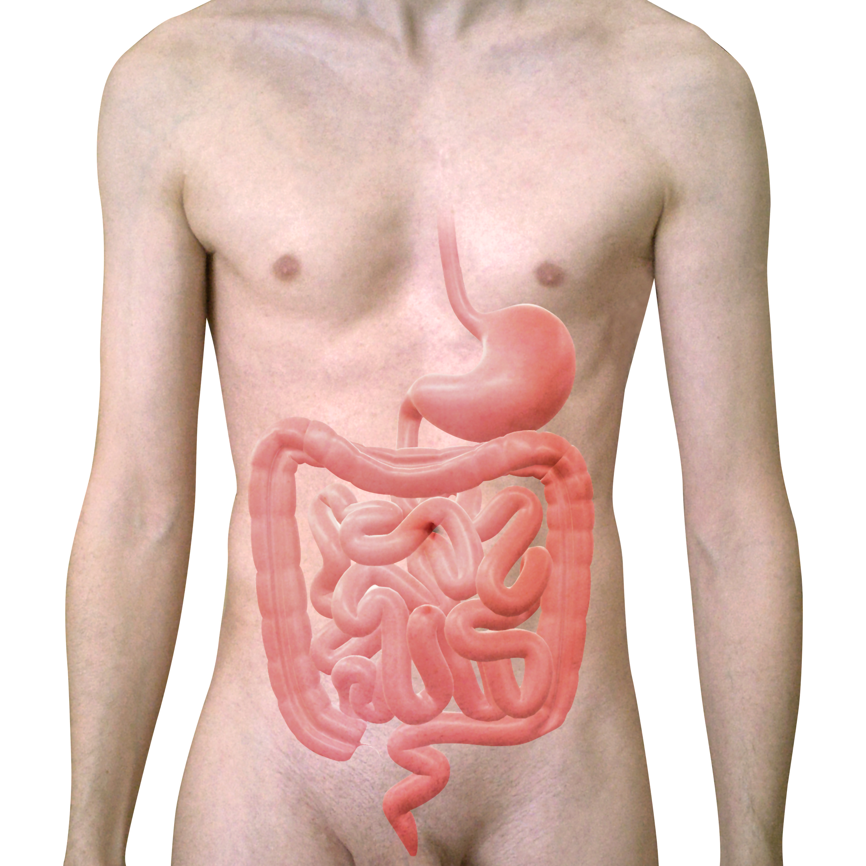
The small intestine is about 6 metres long and roughly 3 centimetres wide, which gives a plain cylinder an internal surface area of about half a square metre. Its actual absorptive surface is around 30 square metres — comparable to a badminton court, and about 60 times more than the tube's shape alone would give.
That multiplication happens by three tricks stacked on top of each other, and it is why essentially everything you absorb is absorbed here.
The three amplifications
Circular folds — permanent ridges running around the inside of the tube, up to 8 millimetres tall. Roughly a threefold increase, and they also make the contents spiral as they move, improving mixing.
Villi — finger-like projections about 1 millimetre tall, giving the lining a velvety texture. Around 20 to 40 per square millimetre. Roughly a tenfold increase.
Microvilli — on the surface of each absorptive cell, about 1 micrometre tall and packed at up to 200,000 per square millimetre. They form the brush border. Roughly a twentyfold increase.
3 \times 10 \times 20 = 600\text{-fold, from a base of about 0.5 m}^2
And the microvilli are not just surface — they are machinery. The brush border membrane carries the final digestive enzymes embedded in it: lactase, sucrase, maltase and peptidases. Digestion of the last few bonds happens at the moment of absorption, on the surface of the very cell that will take the product up.
This is why lactose intolerance is a brush border problem (Chapter 1.2), and why any disease that damages the villi causes multiple deficiencies at once.
Inside a villus

Each villus contains a capillary network and one central lymphatic vessel, the lacteal.
And the two carry different things. Sugars and amino acids enter the capillaries and go to the liver via the portal vein. Fats enter the lacteal and bypass the liver entirely (Chapter 7.8), which is why lymph from the gut turns milky after a fatty meal.
Between the villi are crypts — pits containing stem cells that divide continuously. The entire epithelium is replaced every 3 to 5 days, which makes it one of the fastest-renewing tissues in the body.
That renewal rate explains several things at once. It is why the gut lining is so vulnerable to chemotherapy and radiation (Chapter 1.7). It is why gut damage from infection or coeliac disease can heal completely once the cause is removed. And it is why the gut recovers so well — a lining destroyed by a severe infection is fully rebuilt within a week.
Paneth cells at the bottom of the crypts secrete antimicrobial peptides, protecting the stem cells from the bacteria above.
The three regions
Duodenum (25 cm) — where the bile duct and pancreatic duct empty. Most chemical digestion happens here, and iron and calcium are absorbed here.
It is also where the acid is neutralised. Pancreatic secretion is rich in bicarbonate, and it raises the pH from about 2 to around 6 to 7 — which is required, because every pancreatic enzyme works at neutral pH and is destroyed by acid.
Brunner's glands in the duodenal wall secrete additional alkaline mucus, and they exist only here — a defence against the acid arriving from the stomach.
Jejunum (2.5 m) — the main absorptive region. Most sugars, amino acids and fats are taken up here.
Ileum (3.5 m) — completes absorption, and does two things nowhere else can.
Vitamin B12 absorption, with intrinsic factor, in the terminal ileum only (Chapter 7.1).
Bile salt reabsorption, also in the terminal ileum. About 95 percent of bile salts are recovered and returned to the liver for reuse — the enterohepatic circulation, and it recycles the pool 6 to 10 times a day.
This localisation has direct consequences. Crohn's disease has a particular affinity for the terminal ileum, and surgical removal of that segment produces both B12 deficiency and bile salt malabsorption. The unabsorbed bile salts then reach the colon, where they draw water in and cause diarrhoea — treatable with a drug that binds them.
How each nutrient is absorbed
Carbohydrates. Starch is broken by amylase into shorter chains, then by brush border enzymes into single sugars. Only monosaccharides can be absorbed.
Glucose and galactose enter by SGLT1, the sodium-coupled transporter of Chapter 1.4 — which is why oral rehydration solution works. Fructose enters by a different, facilitated route, which is why some people tolerate it poorly: the capacity is lower and unabsorbed fructose ferments in the colon.
Proteins. Pepsin starts in the stomach; pancreatic trypsin, chymotrypsin and others continue; brush border peptidases finish the job. Amino acids, dipeptides and tripeptides are all absorbed, by several different carriers.
Fats — the most complex, and worth following because it explains several drugs and diseases.
The problem is that fat does not dissolve in water, so enzymes in the watery gut contents cannot reach it. The solution has four steps.
1. Emulsification. Bile salts, made by the liver, are detergents — a water-loving face and a fat-loving face. They break large fat droplets into tiny ones, increasing the surface area available to enzymes by a thousandfold or more. Bile salts do not digest fat; they make it accessible.
2. Digestion. Pancreatic lipase splits triglycerides into fatty acids and monoglycerides.
3. Micelle formation. These products are packaged by bile salts into tiny spheres — micelles — that ferry them to the brush border.
4. Absorption and repackaging. The fatty acids diffuse into the cell, are reassembled into triglycerides, packaged with protein into chylomicrons, and released into the lacteal.
Break any step and fat is malabsorbed, producing steatorrhoea — pale, bulky, greasy, foul-smelling stool that floats.
Missing bile (a blocked bile duct) breaks step 1. Missing lipase (pancreatic disease) breaks step 2. Damaged villi (coeliac disease) break step 4.
And fat-soluble vitamins A, D, E and K travel with the fat, so any fat malabsorption causes their deficiency — which is why people with cystic fibrosis or chronic pancreatitis need supplements of all four.
Orlistat, a weight-loss drug, blocks pancreatic lipase deliberately. It works, and its side effects are precisely the steatorrhoea described above — which is both the reason it is unpopular and, arguably, part of how it modifies behaviour.
Water. About 9 litres pass through the small intestine daily — 2 from drinking and 7 secreted by the gut itself. The small intestine absorbs about 8 litres and the colon most of the rest, leaving about 100 to 200 ml in stool.
Water follows solutes osmotically, which is why absorbing sodium and glucose pulls water with it, and why anything that leaves solute in the lumen causes diarrhoea.
Minerals.
Iron is absorbed in the duodenum, and the body regulates absorption rather than excretion (Chapter 7.1). Absorption is enhanced by vitamin C and by an acidic stomach, and inhibited by tea, coffee, calcium and phytates in whole grains. Practical consequence: take iron tablets with orange juice, not with tea, and separate them from calcium supplements.
Calcium absorption requires vitamin D (Chapter 5.1).
The pancreas as a digestive organ
The endocrine pancreas is Chapter 12.5; here it is the exocrine part.
About 1.5 litres of pancreatic juice a day, containing bicarbonate and the full enzyme set:
- Amylase for starch
- Lipase for fat
- Trypsinogen, chymotrypsinogen and others for protein
- Nucleases for nucleic acids
The protein-digesting enzymes are secreted inactive, for the obvious reason. Trypsinogen is activated in the duodenum by a brush border enzyme, and trypsin then activates everything else — including more trypsinogen.
The pancreas also makes a trypsin inhibitor, as a second line of defence in case any activation happens prematurely inside the gland.
Acute pancreatitis is what happens when those defences fail. Enzymes activate inside the pancreas and it digests itself. The two commonest causes are gallstones — blocking the duct — and alcohol, together accounting for about 80 percent.
It presents as severe upper abdominal pain radiating to the back, with vomiting, and the pain is characteristically eased by leaning forward. Amylase and lipase in the blood are raised, lipase being more specific.
Most cases are mild and settle with fluids and pain relief. A minority become severe, with widespread inflammation, organ failure and a mortality of 20 to 30 percent. Chapter 21.2.
Chronic pancreatitis destroys the gland over years, producing malabsorption and diabetes. Enzyme replacement capsules taken with every meal correct the malabsorption effectively, and getting the dose and timing right transforms these patients' nutrition.
Coeliac disease
An immune reaction to gluten — a protein in wheat, barley and rye — that damages the small intestinal villi.
Not an allergy and not an intolerance. It is an autoimmune disease in which the immune system, having reacted to gluten, attacks the intestinal lining.
The villi flatten, so the surface area collapses, and absorption fails across the board.
It affects around 1 percent of people, and a large proportion remain undiagnosed for years, because the presentation is so variable.
Classic presentation — diarrhoea, weight loss, steatorrhoea, bloating.
But the commoner presentation now is atypical: iron deficiency anaemia that does not respond to iron, unexplained fatigue, osteoporosis at a young age, recurrent mouth ulcers, infertility, or raised liver enzymes. In children, poor growth.
An itchy blistering rash on the elbows, knees and buttocks — dermatitis herpetiformis — is coeliac disease in the skin, and it responds to a gluten-free diet.
Diagnosis is by blood antibody testing followed by biopsy — and the patient must still be eating gluten when tested, because the tests normalise on a gluten-free diet. This is the single most common practical error: someone tries a gluten-free diet, feels better, then gets tested and the result is negative.
Treatment is a strict lifelong gluten-free diet, and the encouraging part is that it works completely. The villi regenerate — remember the 3 to 5 day turnover — and the intestine returns to normal, usually within months. Symptoms resolve, deficiencies correct, and the increased long-term risk of intestinal lymphoma falls back toward normal.
Non-coeliac gluten sensitivity is a separate and genuinely contested entity — people who report symptoms with gluten but have no antibodies and no villous damage. Some of it appears to be a response to fermentable carbohydrates in wheat rather than to gluten itself, and this is an area where honest uncertainty is the correct position.
Other malabsorption
Lactose intolerance — Chapter 1.2. Common, harmless, and manageable: most people with it tolerate small amounts, particularly with meals, and yoghurt and hard cheeses are usually fine because much of the lactose has been fermented or drained off.
Small intestinal bacterial overgrowth (SIBO) — bacteria colonising the small intestine, which is normally relatively sparse. Causes bloating, diarrhoea and B12 deficiency, because the bacteria consume it.
Short bowel syndrome — after extensive surgical removal. The remaining bowel adapts substantially over months to years, with villi lengthening and absorptive capacity increasing, which is a genuinely impressive demonstration of the tissue's plasticity.
What the next page fixes
Everything absorbed here, except fat, goes directly to one organ before reaching the rest of the body. Chapter 9.4 covers the liver — 500 known functions, the only organ that regenerates, and the one whose failure produces the most varied set of symptoms in medicine.