Appearance
5.4 — The Thorax and Shoulder Girdle
Your ribcage has to be rigid enough to protect the heart and lungs from a blow, and mobile enough to expand and contract about 20,000 times a day for eighty years. Those two requirements are met by a design most people never notice: the ribs are not fixed to the sternum by bone. They connect through cartilage, and that cartilage is what lets the whole cage move.
Attached to that cage is the shoulder girdle, which solves an entirely different problem — how to give the arm an enormous range of motion — and pays for it in a way that fills orthopaedic clinics.
The ribs
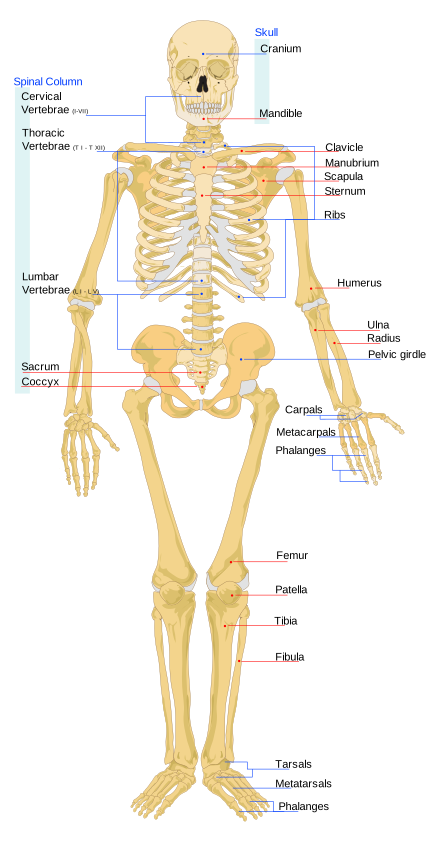
Twelve pairs, all articulating with thoracic vertebrae at the back.
Ribs 1 to 7 are "true" ribs — each connects to the sternum through its own costal cartilage.
Ribs 8 to 10 are "false" ribs — their cartilages join the cartilage above rather than reaching the sternum directly.
Ribs 11 and 12 are "floating" ribs — short, with no anterior attachment at all. They end in the muscle of the abdominal wall.
Each rib slopes downward as it runs forward, so its front end is one to two vertebral levels lower than its back end. This slope is the whole mechanism of breathing.
When the muscles between the ribs contract, they lift the ribs. Because the ribs slope downward, lifting them moves the sternum forward and upward — the "pump handle" motion — increasing the front-to-back diameter of the chest. At the same time the lower ribs swing outward — the "bucket handle" motion — increasing the side-to-side diameter. Both enlarge the chest cavity, which drops the pressure inside and draws air in (Chapter 8.3).
The first rib is the exception in almost every way. It is short, sharply curved, nearly horizontal, and it is the flattest. It is largely hidden under the clavicle, and it is the hardest rib to break. A fractured first rib therefore means very high energy, and it is treated as a marker of severe trauma and a reason to look hard for injury to the great vessels running immediately beneath it.
The sternum
Three parts.
The manubrium at the top, articulating with the clavicles and the first ribs. The body, articulating with ribs 2 to 7. The xiphoid process, a small cartilaginous tip that ossifies in middle age.
The junction between manubrium and body is the sternal angle, a palpable ridge, and it is the single most useful surface landmark in the chest. The second rib attaches exactly there, so it is how you count ribs reliably — you cannot count the first because the clavicle covers it. It also marks, internally, where the trachea divides into the two main bronchi, where the aortic arch begins and ends, and the boundary between upper and lower mediastinum. One ridge you can feel through the shirt tells you where four internal structures are.
The xiphoid matters in resuscitation. Chest compressions are given on the lower half of the sternum, and pressing on the xiphoid itself can break it off and drive it into the liver. The landmark is the centre of the chest, and the xiphoid is what you stay above. Chapter 23.2.
And the sternum is where a marrow sample can be taken, being one of the sites that retains red marrow in adults (Chapter 5.1).
Chest injuries, and the mechanics behind each
Rib fractures are common and mostly heal without intervention. The important consequence is not the bone but the breathing: it hurts to breathe deeply, so the person breathes shallowly, so the base of the lung does not inflate, so secretions collect and pneumonia follows. In the elderly, rib fractures carry a mortality of around 10 to 20 percent for this reason alone. The treatment is therefore adequate pain relief specifically so that the person can breathe deeply and cough — which is the opposite of the instinct to avoid strong painkillers in older patients, and it is a case where undertreating pain kills people.
Flail chest occurs when three or more adjacent ribs are each broken in two places, isolating a segment of chest wall from the rest. That segment then moves paradoxically — sucked inward on inspiration and pushed out on expiration, because it follows the pressure inside the chest rather than the muscles. It is a sign of very high energy trauma, and the underlying lung bruising is usually what threatens life rather than the mechanics.
Pneumothorax — air in the pleural space, collapsing the lung on that side. Because the two pleural cavities are separate (Chapter 4.1), the other lung continues working.
Tension pneumothorax is the emergency version, and it is worth understanding mechanically because recognising it saves lives. A tear acts as a one-way valve: air enters the pleural space with each breath and cannot escape. Pressure rises progressively, collapsing the lung completely, then pushing the mediastinum toward the opposite side, kinking the great veins returning blood to the heart. The person dies of obstructed circulation, not of the lung collapse.
The signs are severe breathlessness, absent breath sounds on one side, a trachea deviated away from that side, distended neck veins and falling blood pressure. The treatment is immediate needle decompression — a large cannula inserted into the chest to let the trapped air out — followed by a chest drain. It is one of the few conditions where treatment must precede an X-ray, because the person will die during the wait.
The clavicle
The collarbone is a slender S-shaped strut connecting the sternum to the scapula.
It is the only bony connection between the arm and the axial skeleton. Everything else attaching the shoulder to the trunk is muscle.
Its job is a strut: it holds the shoulder out from the chest wall, giving the arm room to move. Without it, the shoulder collapses medially and the arm's range drops sharply.
It is the most commonly fractured bone in the body, and the mechanism is nearly always the same: a fall onto an outstretched hand or onto the shoulder, transmitting force along the arm to the clavicle, which fails at its weakest point — the junction of its middle and outer thirds, where its curvature changes. Most heal without surgery in a sling.
It is also the first bone in the body to ossify, at around week 5 of development, and the last to finish, with its inner end not fusing until about 25 — which makes it useful for age estimation in young adults.
And it is the one bone that can be fractured deliberately during birth. In a difficult delivery where the shoulders are stuck, breaking the clavicle reduces the shoulder width enough to deliver the baby. It heals rapidly and completely, and it is a legitimate manoeuvre when the alternative is a brachial plexus injury or worse.
The scapula
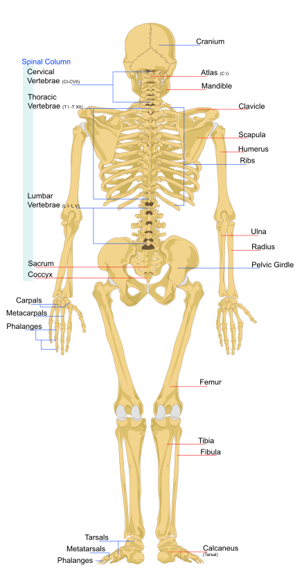
The shoulder blade is a flat triangular bone lying over ribs 2 to 7 at the back.
It is not attached to the ribcage by any joint. It floats on a bed of muscle, and this arrangement — sometimes called the scapulothoracic joint although it is not a true joint — is what allows it to slide up, down, forward, back and rotate.
That mobility contributes about a third of the arm's total range. Raise your arm fully overhead: roughly 120 degrees comes from the shoulder joint itself and roughly 60 degrees from the scapula rotating upward. The two move in a fixed ratio, and when that coordination is disrupted the shoulder does not work properly even if the joint itself is normal — which is why shoulder rehabilitation focuses so heavily on the muscles that control the scapula.
Key features: the spine of the scapula, a prominent ridge you can feel across the upper back; the acromion, its outer end, forming the point of the shoulder and articulating with the clavicle; the coracoid process, a hook projecting forward; and the glenoid cavity, the shallow socket for the humerus.
A scapular fracture is rare and significant. It is well protected by muscle and needs enormous force, so like a first rib fracture it flags severe trauma elsewhere.
The shoulder joint

The shoulder is the most mobile joint in the body and the most frequently dislocated, and those two facts are the same fact.
The humeral head is roughly a hemisphere. The glenoid cavity is a shallow dish about a third of its diameter. The comparison usually made is a golf ball on a tee, and it is fair. Stability is sacrificed almost entirely for range.
Two structures do the stabilising.
The glenoid labrum — a rim of fibrocartilage around the socket, deepening it by about 50 percent and creating a suction effect.
The rotator cuff — four muscles whose tendons blend with the joint capsule and hold the head against the socket actively. Supraspinatus, infraspinatus, teres minor and subscapularis. Chapter 6.5 covers what each does.
Dislocation occurs in about 1 to 2 percent of people at some point. Around 95 percent go forward and downward, because the capsule is weakest there and there is no cuff tendon covering that part.
Two complications follow directly from anatomy. The axillary nerve wraps around the surgical neck of the humerus immediately below the joint, so it can be stretched or compressed — producing numbness over the outer shoulder, which is checked before and after any reduction. And once the labrum and capsule are torn, they often do not heal tight, so recurrence is common — over 80 percent in patients under 20, falling with age. That age dependence is why young patients with a first dislocation are increasingly offered surgical repair rather than sling and physiotherapy.
What the next page fixes
From the shoulder the arm continues into the most versatile manipulator in the animal kingdom — thirty bones per limb, a wrist that permits rotation without twisting the forearm's arteries, and a thumb that can touch every other finger. Chapter 5.5 covers the upper limb bone by bone, including the fractures that are named after people and the reason a scaphoid fracture is the one you must not miss.