Appearance
25.26 — Inside the Hospital: the Operating Theatre and Its Machines
A patient is asleep on a table. Around them, in a room with the air changing twenty times an hour and flowing downward from filters in the ceiling, six people are working. Every one of them is watching a different set of numbers, and almost every object in the room is a regulated medical device.
If your company builds software for hospitals, this is the environment your work eventually reaches. And even if it never does, understanding what happens here is the difference between a person who has read about the industry and a person who can picture it.
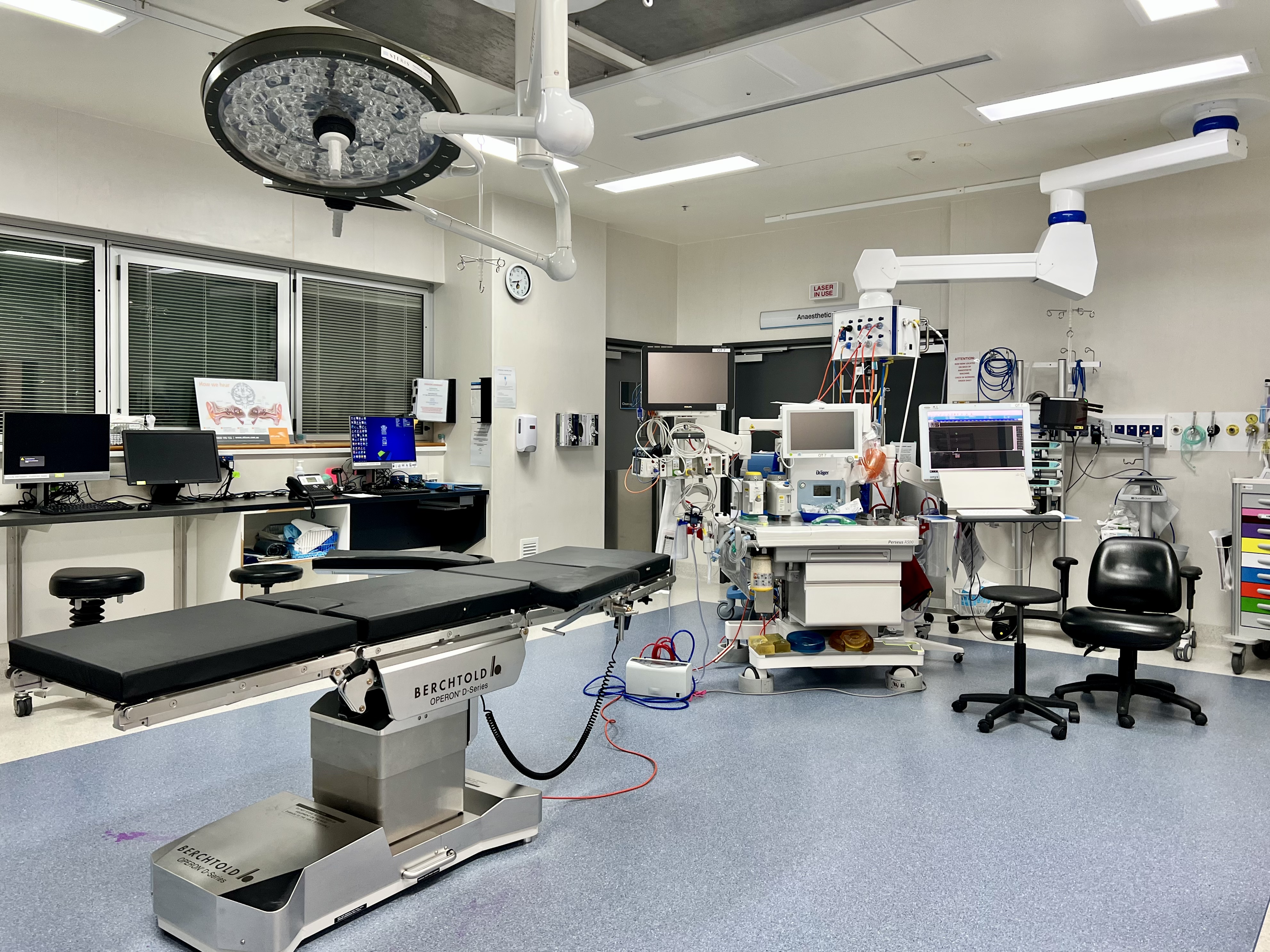
The room itself is a device
Everything about an operating theatre is designed against one enemy: infection.
The air is filtered through high-efficiency filters and exchanged many times an hour, and the room is held at a slightly higher pressure than the spaces around it so that when a door opens, air flows out rather than in. Some theatres for joint replacement use a downward flow of ultra-clean air over the table.
The sterile field is a defined zone — the draped patient, the instrument tables, and the gowned front of the scrubbed team from chest to waist and hands to elbows — and anything that touches something outside it is no longer sterile. The rules are absolute and unembarrassed: a scrubbed person keeps their hands above the waist and in front, and if there is any doubt about whether something was contaminated, it is treated as contaminated.
Instruments are sterilised, most commonly by steam under pressure in an autoclave, typically at 121 degrees for around fifteen minutes or 134 degrees for around three, with the exact cycle depending on the load. Heat-sensitive items go through low-temperature methods such as ethylene oxide gas or hydrogen peroxide plasma. Every load is monitored — with physical readings, chemical indicators that change colour, and biological indicators containing bacterial spores that are cultured afterwards to prove the cycle actually killed things.
And the team is fixed in role. The surgeon and assistants operate. The anaesthetist keeps the patient alive and unconscious. The scrub nurse or technologist manages the instruments and counts them. The circulating nurse moves in the unsterile space, fetching and documenting. For heart surgery, a perfusionist runs the machine that takes over the work of the heart and lungs.
Before anything begins, the team runs a checklist. The World Health Organization's surgical safety checklist has three pauses: before anaesthesia, before the incision, and before the patient leaves the room. It confirms identity, the procedure, the site, allergies, antibiotic timing, expected blood loss, equipment concerns, and — at the end — the instrument and swab counts. The published evaluation of it found substantial reductions in complications and deaths across a range of hospitals, and it costs nothing. It is the clearest example in medicine of a process control outperforming technology.
The machines around the patient
Anaesthesia machine. It delivers a precise mixture of oxygen, air and anaesthetic vapour, ventilates the patient, and scavenges the exhaled gas so the team does not breathe it. Its safety interlocks are the interesting part for an engineer: the machine physically cannot deliver a hypoxic mixture, and its alarms are prioritised so that the critical ones cannot be silenced indefinitely.
Patient monitor. The standard set is heart rhythm by ECG, oxygen saturation by pulse oximeter, blood pressure by cuff or by an arterial line, exhaled carbon dioxide by capnography, and temperature.
Of those, capnography deserves a note, because it is the one that catches disasters first. A waveform of exhaled carbon dioxide confirms that the breathing tube is in the airway rather than the oesophagus, that the lungs are being ventilated, and that blood is circulating to carry carbon dioxide back — so it fails immediately if the tube is misplaced, the circuit disconnects, or the heart stops. It is a single measurement that tests three separate systems at once, which is why it is mandatory.
Electrosurgery unit. High-frequency current is used to cut tissue and seal bleeding vessels. In monopolar mode the current passes through the patient to a large return pad, which is why the pad's placement and contact matter — a poor contact concentrates the return current and causes burns. In bipolar mode the current passes only between the tips of a forceps, which is safer near delicate structures.
Suction, surgical lights, warming devices to keep the patient's temperature up, and infusion pumps delivering drugs and fluids at controlled rates. Modern infusion pumps carry drug libraries with dose limits, and those limits are one of the most effective medication-safety controls in a hospital.
For keyhole surgery, an insufflator fills the abdomen with carbon dioxide to create working space, held at a low, controlled pressure, plus a camera stack and long instruments.
For imaging during the operation, a mobile fluoroscopy unit — the C-arm — takes live X-ray images, and ultrasound is used to guide needles and assess tissue.
And for cardiac surgery, the heart-lung machine, which takes blood from the body, oxygenates it, removes carbon dioxide, controls its temperature, and pumps it back — allowing the heart to be stopped and opened.
Robotic surgery: what it is and what it is not
The dominant system in the world is the da Vinci platform, and the most important thing to understand about it is that it is not autonomous. Every movement of every instrument is commanded by a surgeon in real time.
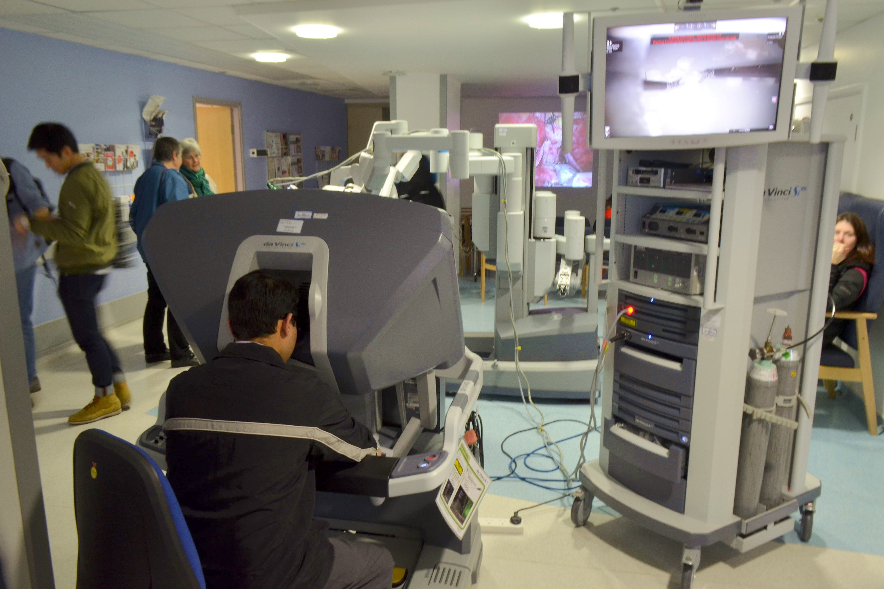
The system has three parts. The console where the surgeon sits, away from the table. The patient cart with three or four arms holding the camera and instruments, docked to ports placed through the abdominal wall. And a vision cart with the light source, image processing and a screen for the rest of the team.
Four capabilities explain why surgeons want it.
Wristed instruments. A laparoscopic instrument is a rigid stick through a fixed point, so its tip cannot articulate. The robot's instruments have a small wrist near the tip with several degrees of freedom, which makes suturing deep in the pelvis practical in a way it is not with straight instruments.
True three-dimensional vision from a stereo camera, rather than a flat image on a wall screen.
Motion scaling and tremor filtering. Hand movement can be scaled down — three centimetres of hand movement becoming one centimetre of instrument movement — and physiological tremor is filtered out.
And ergonomics. The surgeon sits, supported, looking straight ahead, instead of standing for six hours holding instruments above shoulder height. Surgeon fatigue is a real determinant of outcomes in long operations.
Now the honest limitations, because a client's clinical staff will respect you far more for knowing them.
Haptic feedback is limited or absent. The surgeon cannot feel tissue tension the way they can through a laparoscopic instrument, and they compensate by watching visual cues — tissue blanching, suture deformation.
The system must be docked, and if the patient deteriorates and the abdomen must be opened immediately, the cart has to be undocked first — which is a drilled emergency procedure, timed and practised.
It is expensive, in capital cost, per-procedure instrument cost, and theatre time, particularly early in a team's experience.
And the evidence is genuinely mixed by procedure. For some operations — prostate removal is the standard example — robotic assistance became the dominant approach quickly. For others the measurable benefits over conventional keyhole surgery are modest, and the honest summary is that the robot changes what is technically feasible in confined spaces more than it changes outcomes across the board.
Other robotic systems solve different problems. Orthopaedic systems build a three-dimensional plan from a scan and then constrain the surgeon's cutting tool so it cannot stray outside the planned boundary — a physical guardrail rather than teleoperation. Catheter and bronchoscopy systems steer flexible instruments through vessels or airways. And navigation systems track instruments relative to a registered scan, showing the surgeon where the tip is relative to anatomy they cannot see.
Every one of these is a Class II or Class III device under Chapter 25.16, with software safety classification, risk management files, and — increasingly — the cybersecurity obligations described there.
The information systems in and around the theatre
A theatre generates a great deal of data and almost none of it is easy to move.
Scheduling and case management — which patient, which surgeon, which room, what equipment, what implants, and what the turnover time is between cases. Theatre utilisation is one of the highest-value operational metrics in a hospital, because an operating room is among the most expensive spaces in the building.
Anaesthesia information management — automatic capture of monitor readings at short intervals, plus drugs and events, replacing a hand-drawn chart. This is device integration in practice, usually over the hospital messaging standards described in Chapter 25.31.
Implant and instrument traceability — which serial-numbered implant went into which patient, which is exactly the unique device identifier requirement from Chapter 25.16, and which matters enormously on the day a device is recalled.
Video and image capture, increasingly used for training and for automated analysis of surgical steps.
And alarms. A monitored patient generates a great many alarms, most of them not clinically actionable, and staff become desensitised — the phenomenon is called alarm fatigue and it has killed people. Alarm management, meaning intelligent filtering, escalation and routing to the right person, is a serious clinical safety project and a genuinely worthwhile engineering problem.
Why this chapter matters for a software engineer
Three things carry over into every hospital-facing project.
Clinical staff work in an environment where interruption is dangerous. A system that demands attention at the wrong moment will be worked around, and the workaround will be the thing that hurts somebody. Chapter 25.19's point about violations applies exactly here.
Devices are not general-purpose computers. They run for a decade, cannot be patched casually, may not tolerate a network scan, and are regulated such that a change requires assessment. This is why hospital networks segregate medical devices, and why "just update it" is not available.
And every number you display was measured by something with a failure mode. A pulse oximeter reads badly in a cold hand or with nail polish; a blood pressure cuff of the wrong size reads high; an ECG lead that has come loose produces a rhythm that looks fatal. Software that presents these numbers without carrying their quality alongside them is dangerous, and clinicians will judge your product on whether you understood that.
Next: Chapter 25.27, the system that pays for all of this — the American healthcare model, who the players are, and why it works the way it does.