Appearance
2.5 — Mutation, Repair, and What Goes Wrong
Each of your cells suffers somewhere between 10,000 and 100,000 DNA lesions a day. Bases fall off spontaneously. Cytosine slowly converts to uracil by simple chemistry. Oxygen by-products from the mitochondria in Chapter 1.6 attack guanine. Ultraviolet light welds neighbouring bases together. Radiation snaps both strands at once.
Almost all of it is repaired within minutes. A mutation is what is left when repair fails — and you accumulate them at a rate of roughly one to two permanent changes per cell division, plus more from damage.
Most do nothing. Some cause cancer. About 70 of them are new in you, present in neither of your parents, and a handful of those you will pass to your children.
The kinds of mutation
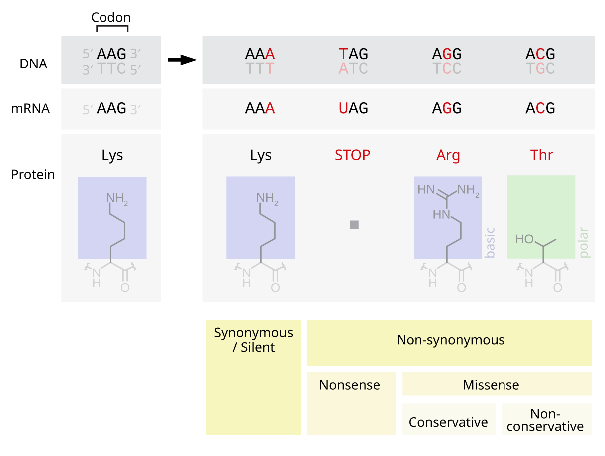
Substitution — one base replaced by another. Three outcomes, and which one you get is largely luck.
- Silent. The new codon specifies the same amino acid, because of the code's redundancy from Chapter 2.3. Nothing changes. Roughly a quarter of random substitutions are silent, and most of those are in the third position of a codon.
- Missense. A different amino acid goes in. The effect ranges from nothing to fatal depending on where it is and what it swaps. Sickle cell disease is a single missense mutation — one A becomes a T, glutamic acid becomes valine, and Chapter 1.3 covered what follows.
- Nonsense. The new codon is a stop codon. Translation halts early and the protein is truncated, usually to a useless fragment. Almost always destructive.
Insertion or deletion. Adding or removing bases. The consequence depends entirely on whether the number is a multiple of three, and this is the single most useful rule in mutation genetics.
Remove three bases and you lose one amino acid; the reading frame is undisturbed and the rest of the protein is normal. Cystic fibrosis's commonest mutation, F508del, is exactly this — three bases gone, one amino acid missing, the rest intact, and yet the protein misfolds (Chapter 1.3).
Remove one or two bases and you get a frameshift. The ribosome reads in fixed triplets from a fixed starting point, so shifting by one changes every codon downstream. The protein after that point is complete nonsense, and a stop codon usually turns up within thirty or so codons by chance. A frameshift near the start of a gene destroys the protein entirely.
The English illustration makes it obvious. Read in threes: THE BIG RED DOG BIT THE MAN. Delete the B from BIG: THE IGR EDD OGB ITT HEM AN. Every word after the deletion is ruined, though nothing after it was touched.
This is why Duchenne and Becker muscular dystrophy are different diseases from the same gene. Both involve deletions in the dystrophin gene. Duchenne deletions shift the frame, so no functional dystrophin is made at all — boys lose the ability to walk by about twelve and historically died in their twenties. Becker deletions remove a whole number of codons, so a shortened but still partly working dystrophin is produced — the disease is far milder and many patients live into their fifties and beyond. Same gene, similar-sized deletions, and the entire difference in outcome is whether the deletion length divides by three. Chapter 21.6.
Repeat expansion. Some genes contain a short sequence repeated many times, and the repeat can grow during replication when the strands slip. Beyond a threshold, disease results. Huntington's disease is the classic: a CAG triplet repeated in the huntingtin gene. Under 27 repeats is normal; 40 or more causes the disease with certainty. And the repeat tends to grow further when passed on, especially through the father, so each generation may develop symptoms earlier than the last — a phenomenon called anticipation, which looked mysterious for decades and is a direct mechanical consequence of slippage. Fragile X syndrome and myotonic dystrophy work the same way.
Larger events — whole chunks duplicated, deleted, inverted, or moved between chromosomes. Chapter 2.7 covers the chromosomal scale.
Where mutations come from
Spontaneous chemistry is the largest source and needs no external agent. Around 10,000 purine bases fall off the backbone of each cell's DNA every day. Cytosine spontaneously converts to uracil a few hundred times a day per cell.
That last one explains why DNA uses thymine and RNA uses uracil. Thymine is chemically just uracil with a methyl group added. If DNA used uracil normally, a cytosine that had converted to uracil would be indistinguishable from a legitimate base and would never be repaired. Because DNA uses thymine, any uracil found in DNA is definitionally an error, and a dedicated enzyme cuts it out. The apparently wasteful extra methyl group is an error-detection scheme, and the same logic appears in every checksum in Volume I, Chapter 1.8.
Replication errors — about three per genome copy, after all three proofreading layers (Chapter 2.2).
Reactive oxygen species from your own mitochondria. This is the price of aerobic metabolism.
Radiation. Ultraviolet light, mostly UVB, causes adjacent thymines to bond to each other, forming a thymine dimer that kinks the helix and blocks replication. X-rays and gamma rays cause double-strand breaks, which are the most dangerous lesion of all because there is no intact strand to copy from.

Chemicals. Benzopyrene from tobacco smoke and grilled meat inserts itself into DNA and causes substitutions — and it has a preference for specific positions in the p53 gene that match exactly the mutations found in lung tumours from smokers, which is about as direct a chain of evidence from cause to disease as toxicology gets. Aflatoxin from mould on badly stored groundnuts and maize causes a very specific p53 mutation and liver cancer. Nitrosamines in processed meat, and aromatic amines in industrial dyes, are others.
Viruses. Some insert their DNA into yours, disrupting whatever gene they land in. Human papillomavirus does this and causes cervical cancer (Chapter 19.5).
The repair systems
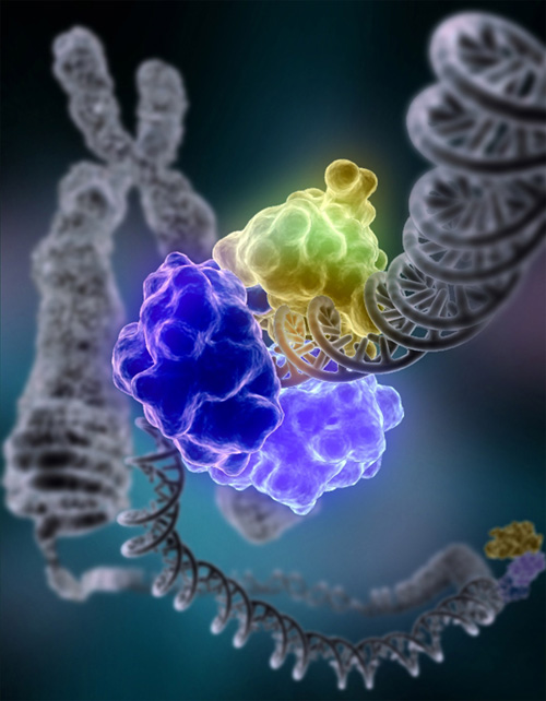
Five systems, each with a disease attached when it fails. The disease is the proof that the system matters.
Base excision repair. A glycosylase enzyme recognises one chemically wrong base — a uracil, an oxidised guanine — flips it out of the helix, and cuts it off. The gap is filled and sealed. Handles the largest volume of damage.
Nucleotide excision repair. For bulky lesions that distort the helix, such as thymine dimers. A stretch of about 25 to 30 nucleotides around the damage is removed and resynthesised from the other strand.
When this fails: xeroderma pigmentosum. People with this inherited condition cannot repair ultraviolet damage. They develop severe sunburn from minimal exposure, and skin cancers begin appearing in early childhood — the risk is over a thousand times normal, with a median first skin cancer around age eight. Affected children are sometimes called "children of the night" because avoiding daylight entirely is the only effective protection. It is the clearest demonstration available that ordinary sunlight is genuinely mutagenic and that you are protected only because your repair works.
Mismatch repair. Covered in Chapter 2.2; Lynch syndrome when it fails.
Homologous recombination. For double-strand breaks, and it is the accurate solution. It uses the sister chromatid — an identical copy made during replication — as a template, so the break is rebuilt correctly. BRCA1 and BRCA2 are central to this pathway, and that is why faults in them cause cancer: breaks that should have been repaired accurately are instead patched by the sloppy alternative below.
Non-homologous end joining. The fast, inaccurate fallback for double-strand breaks: the two ends are simply stuck back together, usually losing a few bases. Available at any time, including when no sister chromatid exists, and used constantly. It is deliberately imprecise in one place — the immune system uses a controlled version of it to generate antibody diversity (Chapter 13.3).
PARP inhibitors: turning a repair defect into a weapon
This deserves its own section, because it is the best current example of understanding a mechanism and then exploiting it.
Cells with faulty BRCA cannot do homologous recombination. They survive because a different system, single-strand break repair, catches most damage before it ever becomes a double-strand break. An enzyme called PARP is central to that system.
Block PARP, and single-strand breaks go unrepaired. In a normal cell that is manageable — the breaks become double-strand breaks at the replication fork, and homologous recombination fixes them. In a BRCA-mutant cell there is no homologous recombination, so the breaks accumulate and the cell dies.
The tumour has BRCA mutations; the patient's healthy cells still have one working copy. So the drug kills the cancer and largely spares the person. This principle is called synthetic lethality: two defects that are each survivable alone are lethal together. Olaparib and its relatives are now standard in BRCA-associated ovarian, breast, pancreatic and prostate cancer. Chapter 19.7.
Germline versus somatic: the distinction that decides everything
A somatic mutation happens in an ordinary body cell. It affects that cell and its descendants and dies with you. Essentially all cancer is somatic.
A germline mutation is in an egg or sperm, so it ends up in every cell of the resulting child and is passed on. All inherited disease is germline.
This distinction is the whole difference between "I have cancer" and "my family has a cancer risk", and it is why a tumour is sometimes sequenced separately from a blood sample: the tumour shows what went wrong in that cancer, the blood shows what was inherited and therefore what relatives should be told about.
New mutations are commoner than people expect. Each child carries roughly 70 new mutations present in neither parent, most of them harmless. Around 80 percent come from the father, and the number rises with paternal age — about 25 new mutations from a 20-year-old father, around 65 from a 40-year-old. The reason is mechanical: sperm precursor cells keep dividing throughout life, and every division risks a copying error, while eggs stop dividing before birth. This is why some dominant conditions such as achondroplasia appear in children of unaffected parents, and why paternal age is associated with a modestly raised risk of certain conditions.
What you can actually do about it
Most mutation is unavoidable chemistry. Some is not, and the avoidable share is worth naming plainly.
Do not smoke. Tobacco smoke contains at least 70 established carcinogens, and the mutation load in a smoker's lung cells is measurably higher — around 150 extra mutations per lung cell per year of smoking a pack a day.
Protect against ultraviolet light. Sunburn is DNA damage, and the peeling described in Chapter 1.8 is the cells that could not be repaired killing themselves. Sunbeds are a Group 1 carcinogen, the same classification as tobacco and asbestos.
Do not eat visibly mouldy grain or nuts, because of aflatoxin.
Take radiation exposure seriously but proportionately. A chest X-ray delivers about 0.1 millisieverts, roughly ten days of natural background; a CT of the abdomen delivers around 8 to 10 millisieverts, roughly three years' worth. Neither is a reason to refuse a scan you need — the risk of missing a diagnosis is almost always larger — but it is a reason not to request scans that will not change the decision. Chapter 16.6.
And be honest about what antioxidant supplements do, which on the current evidence is not much. Large trials of beta-carotene supplements in smokers found more lung cancer in the supplemented group, not less. Chapter 24.2.
What the next page fixes
This page treated genes one at a time. Inheritance does not work one gene at a time — genes come in pairs on chromosomes, one member from each parent, and the patterns that produces are the whole of classical genetics. Chapter 2.6 covers Mendel, dominance and recession, and the pedigree patterns a doctor uses to work out whether a condition in a family will appear in the next generation.