Appearance
11.1 — The Neuron and the Action Potential
Your brain contains about 86 billion neurons and roughly the same number of glial cells. A single neuron may connect to 10,000 others, giving something in the region of 100 trillion connections. The whole thing runs on about 20 watts — less than a dim light bulb, and a fraction of what any computer performing comparable tasks consumes.
It works by moving ions across membranes. The signal is not electricity in the wire sense — no electrons flow along an axon — it is a wave of ion movement travelling along a membrane, and understanding that is what makes every neurological drug, poison and disease make sense.
The neuron
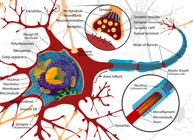
Dendrites — branching processes receiving signals. A single neuron may have thousands, and they are covered in small protrusions called spines where synapses form. The number and shape of those spines change with learning, which is one of the physical substrates of memory.
Cell body (soma) — contains the nucleus and does the housekeeping. It also sums the incoming signals.
Axon hillock — where the axon leaves the cell body. This is the decision point: it has the highest density of voltage-gated sodium channels, so it is where an action potential is initiated.
Axon — the output cable. One per neuron, though it may branch. Length ranges from a fraction of a millimetre to over a metre.
Myelin sheath — insulating wraps of membrane, produced by oligodendrocytes in the brain and cord and by Schwann cells in peripheral nerves (Chapter 4.2). Up to 100 layers of membrane.
Nodes of Ranvier — gaps in the myelin, about 1 micrometre wide, every 0.2 to 2 millimetres.
Axon terminals — the endings that form synapses.
Neurons come in three functional types: sensory (carrying information in), motor (carrying commands out), and interneurons — which are the overwhelming majority, and are the ones doing the processing between the two.
The resting membrane potential
A resting neuron sits at about −70 millivolts, meaning the inside is 70 mV negative relative to outside.
Three things produce it.
The sodium–potassium pump (Chapter 1.4) creates the gradients: potassium high inside, sodium high outside. And it is electrogenic, moving three positive charges out for every two in, contributing a few millivolts directly.
Potassium leak channels are open at rest, and sodium channels are largely closed. So the membrane is far more permeable to potassium than to sodium.
Potassium therefore leaks out down its concentration gradient — and each potassium leaving takes a positive charge with it, leaving the inside more negative.
As the inside becomes more negative, it starts pulling potassium back in electrically. Equilibrium is reached when the concentration gradient pushing potassium out exactly balances the electrical gradient pulling it back.
That equilibrium voltage is given by the Nernst equation:
E_K = \frac{61}{z} \log_{10}\frac{[K^+]_{out}}{[K^+]_{in}}
Putting in the numbers — 4 mmol/L outside, 140 inside, and a charge of +1:
E_K = 61 \times \log_{10}\frac{4}{140} = 61 \times (-1.54) \approx -94\ \text{mV}
So potassium alone would give −94 mV. The actual resting potential is −70 mV, and the difference is the small sodium leak pulling it slightly positive.
And this equation explains directly why potassium disorders are so dangerous (Chapter 10.3). Raise external potassium and the log term shrinks, so the resting potential becomes less negative — the cell is partly depolarised and closer to firing. Initially this makes cells more excitable; push it further and the sodium channels become inactivated and nothing can fire at all, which is why severe hyperkalaemia stops the heart.
The action potential
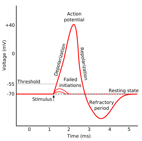
Five phases.
1. Resting. −70 mV. Sodium channels closed, potassium leak channels open.
2. Threshold. Incoming signals depolarise the membrane. At about −55 mV, voltage-gated sodium channels open.
3. Depolarisation. Sodium rushes in, making the inside more positive, which opens more sodium channels — positive feedback (Chapter 4.7). This is why the action potential is all-or-nothing: once threshold is crossed, the process runs to completion regardless of how large the original stimulus was.
The membrane peaks at about +30 mV.
4. Repolarisation. Two things happen together. Sodium channels inactivate automatically after about a millisecond — a separate gate swings shut and cannot be reopened until the membrane repolarises. And voltage-gated potassium channels, which open more slowly, are now open, so potassium floods out.
5. Hyperpolarisation. The potassium channels are slow to close, so a little too much potassium leaves and the membrane dips to about −80 mV before settling back.
The whole event lasts about 1 to 2 milliseconds.
And a point worth making because it surprises people: very few ions actually move. The number of sodium ions entering during a single action potential changes the internal concentration by well under a thousandth of a percent. A neuron could fire thousands of times without the pump running at all — the pump maintains the gradient over the long term, not beat by beat.
The refractory period
Absolute refractory period — during the action potential and briefly after, no stimulus of any strength can trigger another, because the sodium channel inactivation gates are shut.
Relative refractory period — a stronger than normal stimulus can trigger one.
Two consequences follow, and both are essential.
The impulse can only travel forward. The membrane behind it is refractory, so it cannot propagate backward.
And it sets a maximum firing rate, of around 500 to 1,000 impulses per second.
All-or-nothing, and how intensity is encoded
Every action potential in a given neuron is identical in size. A painful stimulus does not produce a bigger impulse than a gentle one.
So intensity is encoded two ways.
Frequency — a stronger stimulus produces more impulses per second. Recruitment — a stronger stimulus activates more neurons.
This is exactly the same solution as a digital signal, and for the same reason: a signal that is always the same size cannot degrade with distance, so it arrives at the far end of a metre-long axon as strong as it started.
Conduction
In an unmyelinated axon, the current spreading from an active region depolarises the adjacent membrane to threshold, which fires, and so on. Continuous conduction, and it is slow — 0.5 to 2 metres per second.
Speed increases with diameter, which is why the squid giant axon — nearly a millimetre thick, used for a rapid escape reflex — was where the action potential was first worked out, by Hodgkin and Huxley in 1952. They shared the Nobel Prize in 1963.
But diameter is an expensive way to gain speed. To double conduction velocity you need roughly four times the cross-sectional area. A human nerve built to squid principles would be impossibly bulky.
Myelination is the alternative.
The myelin insulates the axon so current cannot leak out, and there are no voltage-gated channels under the sheath. Channels are concentrated at the nodes.
So the impulse jumps from node to node — saltatory conduction, from the Latin saltare, to leap. The current spreads passively and almost instantly along the insulated internode, and the action potential is regenerated at each node.
Speed: up to 120 metres per second — about 430 km/h — in a myelinated fibre only 20 micrometres thick.
And it is more efficient as well as faster, because ion movement only happens at the nodes, so far less pumping is needed afterwards.
This is why demyelinating diseases are so disabling. In multiple sclerosis, patches of myelin in the brain and cord are destroyed (Chapter 20.5). Conduction slows dramatically or fails entirely at the affected patch, and the effect depends on which tract is involved — optic nerve, sensory pathway, motor pathway.
In Guillain–Barré syndrome, peripheral myelin is attacked, usually after an infection, causing rapidly ascending weakness that can reach the respiratory muscles within days. It is largely reversible, because peripheral myelin regenerates — which is exactly the difference from multiple sclerosis, where central myelin does not.
Nerve fibre types
Fibres are classified by diameter and myelination, and the classification maps directly onto clinical experience.
| Type | Diameter | Speed | Carries |
|---|---|---|---|
| A-alpha | 13–20 µm | 80–120 m/s | Motor to muscle, position sense |
| A-beta | 6–12 µm | 35–75 m/s | Touch, pressure, vibration |
| A-delta | 1–5 µm | 5–30 m/s | Sharp pain, cold |
| C | 0.2–1.5 µm | 0.5–2 m/s | Dull pain, warmth, itch — unmyelinated |
The two pain fibres explain the two-phase experience of injury, which everyone has had and few have had explained.
Stub your toe and you feel a sharp, well-localised pain immediately — that is A-delta, arriving at 20 m/s. A second or so later comes the deep, aching, poorly localised pain that lasts — that is C fibre, arriving at 1 m/s.
From a toe, over about a metre, the delay is roughly one second, and you can genuinely time it.
And this explains local anaesthetics. Small unmyelinated fibres are blocked first, so the sequence of loss is pain, then temperature, then touch, then pressure, then motor function. This is why a dental anaesthetic abolishes pain while you can still feel pressure and move your jaw, and why patients report feeling the dentist pushing but not hurting.
It also explains why pressure on a nerve — sleeping on your arm — causes numbness before weakness, and why the recovery order reverses.
Channels as drug targets
Voltage-gated sodium channels are blocked by local anaesthetics — lidocaine, bupivacaine — which enter the channel from inside and plug it.
And they are preferentially blocked in rapidly firing neurons, because the channel must open for the drug to enter. This is "use-dependent block", and it is genuinely useful: an inflamed, rapidly firing nerve is blocked more effectively than a quiet one.
The same channels are the target of several anticonvulsants — phenytoin, carbamazepine, lamotrigine — which stabilise them in the inactivated state, so rapidly firing neurons are suppressed while normal ones are barely affected (Chapter 20.2).
And they are the target of some of the most potent natural toxins known. Tetrodotoxin, from pufferfish, blocks them from outside with extraordinary potency — the lethal dose is measured in micrograms, and there is no antidote. Death is from respiratory paralysis, with consciousness preserved, and treatment is supportive ventilation until the toxin clears. Saxitoxin, from the algae that cause red tides, acts identically and causes paralytic shellfish poisoning.
Potassium channels are the target of some antiarrhythmics — and their unintended blockade is the mechanism of drug-induced long QT syndrome (Chapter 7.3).
Calcium channels control transmitter release, and blocking them is how gabapentin and pregabalin work in neuropathic pain.
Glial cells
Roughly as numerous as neurons, and doing far more than support (Chapter 4.2).
Astrocytes regulate the extracellular environment, buffer potassium released by firing neurons, supply neurons with lactate as fuel, and form part of the blood–brain barrier. They also modulate synapses directly, which is a relatively recent finding that has changed how the brain is thought about.
Oligodendrocytes myelinate central axons — each one myelinates up to 50 different axons.
Schwann cells myelinate peripheral axons — one cell per internode of one axon.
And that structural difference explains the regeneration difference. A cut peripheral nerve can regrow at about 1 mm per day, because Schwann cells form a guiding tube and actively support regrowth. A cut central axon does not, because oligodendrocytes and the scar formed by astrocytes actively inhibit it. This is the fundamental reason spinal cord injury is permanent and a cut nerve in the arm is not.
Microglia are the brain's immune cells, and they also prune synapses during development and learning.
What the next page fixes
The action potential reaches the end of the axon and stops — there is a gap. Chapter 11.2 covers the synapse: how the signal crosses, the dozen or so chemicals that carry it, and why almost every drug that changes how you feel acts at that gap.