Appearance
5.5 — The Upper Limb
Each arm contains 30 bones — one in the upper arm, two in the forearm, eight in the wrist and nineteen in the hand. More than half the bones in your entire body are in your hands and feet, and the reason is that fine control requires many short segments rather than few long ones.

The humerus
The single bone of the upper arm, and the longest bone in the upper limb.
Key features and what runs past each one:
The head — a hemisphere articulating with the glenoid (Chapter 5.4).
The surgical neck — a slight narrowing just below the head, so named because it is where fractures happen and surgeons operate. The axillary nerve and the circumflex humeral vessels wrap around it, so a fracture here can cause numbness over the outer shoulder and weakness of the deltoid.
The radial groove — a shallow spiral running down the back of the shaft. The radial nerve lies directly in it. A mid-shaft humeral fracture can therefore injure the radial nerve, and the result is wrist drop: the person cannot extend the wrist or fingers, and the hand hangs limply. It is one of the classic nerve injuries and it is checked for specifically in any humeral fracture.
The medial epicondyle — a bony prominence on the inner side of the elbow. The ulnar nerve runs in a groove behind it, immediately under the skin. This is the "funny bone", and striking it produces the characteristic electric shock down to the little finger — which is not funny and is not a bone but a nerve being hit against bone. Prolonged pressure here, such as leaning on the elbow, causes the same symptoms chronically.
The distal humerus flares into two articular surfaces: a spool-shaped trochlea for the ulna and a rounded capitulum for the radius.
In children, a supracondylar fracture — just above the elbow — is one of the most consequential injuries in paediatric orthopaedics. The displaced fragment can compress the brachial artery and the median nerve running immediately in front of it. Missed, it causes Volkmann's ischaemic contracture, in which the forearm muscles die from lack of blood supply and are replaced by scar, permanently clawing the hand. This is why the pulse and nerve function are checked and documented before and after any manipulation, and why these fractures are treated urgently rather than overnight.
The radius and ulna
Two bones, and the arrangement is the reason your hand can turn over.
The ulna is on the little-finger side. It is the main bone at the elbow — its hook-shaped upper end wraps around the humerus, and the olecranon is the point of the elbow you lean on.
The radius is on the thumb side. It is the main bone at the wrist — it carries most of the load from the hand.
So the load transfers diagonally across the forearm, from the radius at the wrist to the ulna at the elbow, through an interosseous membrane between them.
Pronation and supination. In supination the two bones lie parallel. In pronation the radius rotates over the ulna, crossing it, while the ulna stays put. The hand follows the radius.
This is a much better design than rotating at the shoulder, and the reason is what runs through the arm: rotating the whole limb 180 degrees would twist the brachial artery and the nerves. Rotating one bone over another lets the hand turn without twisting the plumbing.
And it explains a common childhood injury. In a young child the head of the radius is held in place by a ring-shaped ligament that has not yet fully developed its grip. A sudden pull on the arm — lifting a child by one hand, or a tug when they resist — can slip the radial head out from under it. This is "pulled elbow" or nursemaid's elbow. The child stops using the arm and holds it slightly bent and pronated. Reduction is a simple manoeuvre taking seconds, after which the child usually uses the arm again within minutes, and it is one of the most satisfying interventions in a paediatric emergency department.
Two named fractures
Colles' fracture — a fracture of the distal radius with the fragment displaced backward, producing the "dinner fork" deformity when viewed from the side. The commonest fracture in people over 50, and the mechanism is a fall onto an outstretched hand. It is so strongly associated with osteoporosis that a Colles' fracture in a postmenopausal woman is treated as a reason to assess bone density, because it often precedes a hip fracture by several years and is therefore a warning that can be acted on. Chapter 5.9.
Smith's fracture is the reverse — the fragment displaced forward, from a fall onto the back of the flexed hand.
The wrist
Eight carpal bones in two rows of four.
Proximal row, thumb side to little finger: scaphoid, lunate, triquetrum, pisiform. Distal row: trapezium, trapezoid, capitate, hamate.
The scaphoid deserves its own section because it is the fracture that must not be missed.
It bridges both rows and takes the force from a fall onto an outstretched hand, so it is the most commonly fractured carpal bone. The problem is its blood supply, which enters at its far end and runs backward through the bone toward the wrist.
So a fracture across the middle cuts off the blood supply to the proximal half. That fragment then dies — avascular necrosis — and the fracture never heals, leading to collapse and early arthritis of the whole wrist in a young person.
And it is easy to miss. The pain is in the "anatomical snuffbox", the hollow at the base of the thumb, and the fracture is frequently invisible on the initial X-ray, becoming visible only after 10 to 14 days when bone resorption at the fracture edges widens the line.
Therefore the rule: tenderness in the anatomical snuffbox after a fall onto the hand is treated as a scaphoid fracture even with a normal X-ray. The wrist is immobilised and re-imaged, or an MRI or CT is done immediately. Treating a sprain unnecessarily for two weeks costs very little; missing a scaphoid fracture costs a wrist. It is one of the standard teaching examples of managing a patient rather than an image.
The hamate has a hook projecting into the palm, and it can be fractured by the handle of a golf club or racquet — and the ulnar nerve runs beside it, so the fracture can cause weakness of the small hand muscles.
The carpal tunnel is the space between the carpal bones and a tough ligament roofing them over. Nine tendons and the median nerve pass through it. The tunnel cannot expand, so any swelling compresses the nerve — giving numbness and tingling in the thumb, index, middle and half the ring finger, characteristically worse at night, and later weakness of the thumb muscles. Chapter 11.8.
The hand
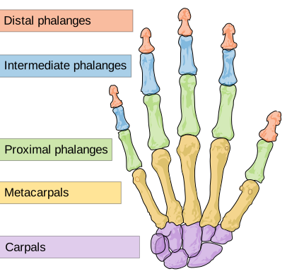
Five metacarpals form the palm, numbered from the thumb. Fourteen phalanges — three in each finger (proximal, middle, distal), two in the thumb.
The thumb is the reason the human hand is different, and the difference is one joint. The joint between the thumb's metacarpal and the trapezium is saddle-shaped, permitting movement in two planes plus rotation. That is what allows opposition — bringing the thumb pad to touch the pad of any other finger.
Precision grip (thumb pad to fingertip, for a pen or a needle) and power grip (all fingers wrapped, thumb reinforcing, for a hammer) both depend on it. Other primates have partial opposition; the human thumb is longer relative to the fingers and its musculature is stronger, which is what makes the precision grip precise.
And that saddle joint's mobility costs it wear. Osteoarthritis at the base of the thumb is one of the commonest sites of arthritis in the hand, particularly in women, and it is disabling out of proportion to its size because almost every grip uses it.
Two fracture patterns worth knowing.
Boxer's fracture — the neck of the fifth metacarpal, from punching with a closed fist. The name is slightly wrong: trained boxers strike with the index and middle knuckles, and it is the untrained punch, landing on the little-finger side, that causes it. Considerable angulation is tolerated because the little finger's function is forgiving.
Bennett's fracture — through the base of the thumb metacarpal into the joint. Fractures involving a joint surface behave differently from shaft fractures — the surface must be restored precisely, because a step of even a millimetre leads to arthritis, so these usually need fixing.
And a hand wound over a knuckle after a punch is treated as an infected human bite until proven otherwise. The mechanism is that the knuckle strikes a tooth, which penetrates the joint capsule; when the hand opens, the puncture in the tendon and the puncture in the skin no longer line up, sealing bacteria inside the joint. Human mouth flora is nasty, and these become septic joints. They get antibiotics and surgical washout, not a plaster.
The nerve supply, and the three signs
Three nerves supply the hand, and each has a characteristic failure pattern that can be tested in seconds. This is one of the highest-value pieces of clinical anatomy in the body, because it lets you localise an injury without any equipment.
Median nerve — sensation to the thumb, index, middle and half the ring finger on the palm side; motor to most thumb muscles. Test: make an "OK" sign. A median nerve problem cannot flex the tip of the thumb and index finger, so the circle collapses into a pinch.
Ulnar nerve — sensation to the little finger and half the ring finger; motor to nearly all the small muscles between the fingers. Test: spread the fingers apart against resistance, or hold a piece of paper between straightened thumb and index finger. Ulnar palsy produces a claw hand.
Radial nerve — sensation to the back of the hand over the thumb side; motor to all the wrist and finger extensors. Test: extend the wrist against resistance. Radial palsy gives wrist drop.
These three tests take about fifteen seconds and are done on every arm injury, because a nerve injury found immediately can be repaired and one found at follow-up often cannot.
What the next page fixes
The upper limb is built for mobility and manipulation, hanging from a girdle barely attached to the trunk. The lower limb is built for the opposite — carrying weight and transmitting force — and its girdle is fused solidly to the spine. Chapter 5.6 covers the pelvis, which is also the one part of the skeleton where male and female anatomy differ profoundly, for reasons that go directly back to Chapter 3.6.