Appearance
11.8 — Peripheral Nerves and Dermatomes
Shingles produces a rash in a band that stops dead at the midline of the body, as though drawn with a ruler. That band is a dermatome — the strip of skin supplied by one spinal nerve — and it is a direct record of the segment the embryo was built from (Chapter 4.4).
The segmental map is why a pattern of numbness tells you where the problem is, and it is one of the most practical pieces of anatomy in this volume.
The dermatome map
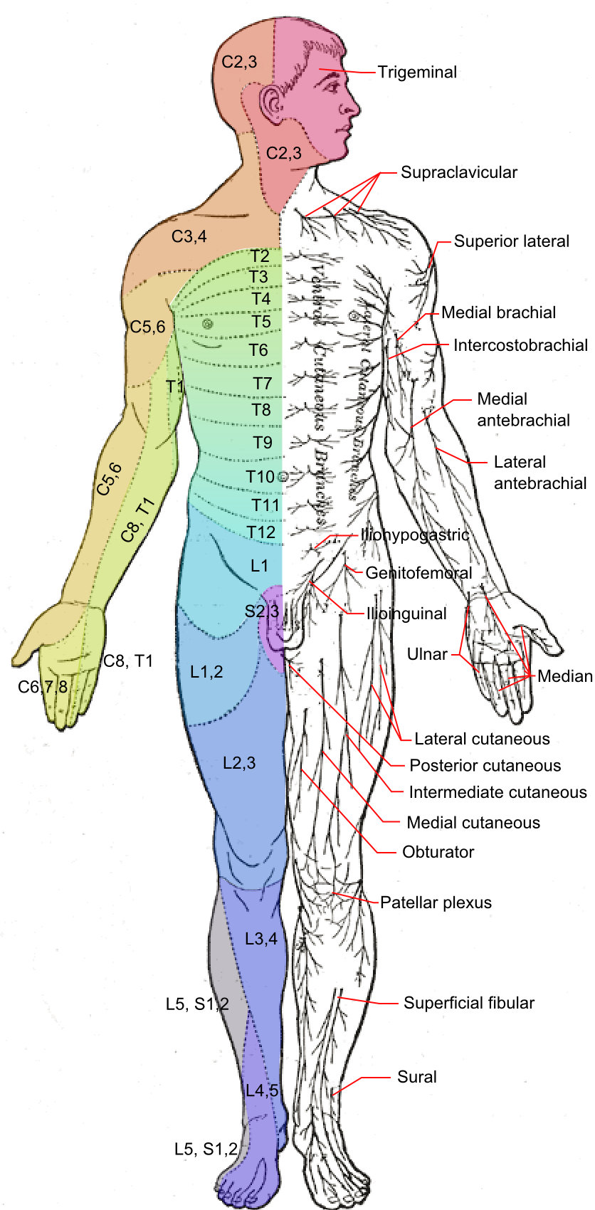
On the trunk the bands are neat horizontal stripes, because the trunk never rotated during development.
On the limbs they look scrambled, and the reason is that the limbs grew out as buds and then rotated (Chapter 4.5). The map is not random — it is a straightforward segmental sequence that has been twisted.
A small number of landmarks are worth memorising, because they let you localise a spinal level anywhere on the body without a chart.
| Level | Landmark |
|---|---|
| C2 | Back of the head |
| C4 | Over the shoulders |
| C6 | Thumb |
| C7 | Middle finger |
| C8 | Little finger |
| T4 | Nipple line |
| T10 | Navel |
| L1 | Groin |
| L4 | Inner shin, and the big toe's inner side |
| L5 | Top of the foot, big toe |
| S1 | Little toe, sole, heel |
| S2–S5 | Saddle area |
Two memory aids that work. T4 at the teat, T10 at the belly button. And on the hand, C6, C7, C8 run thumb, middle, little — in order.
Adjacent dermatomes overlap substantially, which is why damage to a single root causes altered sensation rather than complete numbness. You need two adjacent roots destroyed to produce a genuinely anaesthetic band, and that overlap is a form of redundancy.
And this is exactly why the shingles rash is so distinctive. The virus reactivates in a single dorsal root ganglion (Chapter 11.3) and travels down that one nerve. The rash stops at the midline because a spinal nerve supplies one side only, and there is no crossing.
Shingles is worth two more sentences of practical detail. Antiviral treatment started within 72 hours reduces the severity and the risk of the pain persisting afterwards — post-herpetic neuralgia, which can last months to years. And shingles involving the tip of the nose indicates the nasociliary branch is affected, which means the eye is at risk and needs same-day ophthalmology review. A vaccine substantially reduces both shingles and post-herpetic neuralgia in older adults, and it is one of the more worthwhile vaccines available in later life.
Myotomes
The muscle equivalent — the muscles supplied by one spinal root.
Each muscle usually receives two or three roots, so a single root lesion causes weakness rather than paralysis.
| Movement | Root |
|---|---|
| Shoulder abduction | C5 |
| Elbow flexion | C5, C6 |
| Wrist extension | C6 |
| Elbow extension | C7 |
| Finger flexion | C8 |
| Finger spread | T1 |
| Hip flexion | L1, L2 |
| Knee extension | L3, L4 |
| Ankle dorsiflexion | L4, L5 |
| Big toe extension | L5 |
| Ankle plantarflexion | S1 |
Combined with the reflexes from Chapter 11.3, a full segmental picture can be built in a couple of minutes.
Two maps, one skin — and why the distinction matters
This is the central practical idea of the chapter.
A dermatome is the skin supplied by one spinal root.A peripheral nerve territory is the skin supplied by one named nerve — which is formed from fibres of several roots mixed together in a plexus.
So numbness in a band pattern means a root problem. Numbness in a nerve's territory means a problem in that nerve.
Example: numbness of the thumb.
If the whole radial side of the forearm and the thumb are affected, in a strip, with weak elbow flexion and a reduced biceps reflex — that is a C6 root problem, usually a cervical disc.
If only the thumb, index, middle and half the ring finger are affected on the palm side, worse at night, with weak thumb muscles — that is the median nerve at the wrist, carpal tunnel syndrome.
Different maps, different levels, entirely different treatment, distinguished by where the numbness stops.
The plexuses
Where roots are mixed and redistributed into named nerves.
The brachial plexus — C5 to T1, supplying the arm. A complex rearrangement in the neck and armpit, and its structure is worth knowing only to the extent that it explains the injury patterns.
Erb's palsy — damage to the upper roots, C5 and C6. Classically from excessive traction on the neck, either during a difficult delivery when the shoulder is stuck, or from a fall onto the shoulder.
The arm hangs limply, rotated inward, with the forearm pronated — the "waiter's tip" position. Shoulder abduction and elbow flexion are lost, which is exactly what C5 and C6 supply.
Klumpke's palsy — damage to the lower roots, C8 and T1, from traction on an abducted arm. The small muscles of the hand are paralysed, giving a claw hand, and there may be a drooping eyelid and small pupil on that side, because sympathetic fibres run with T1.
Thoracic outlet syndrome — compression of the plexus or the vessels between the neck muscles and the first rib (Chapter 6.3).
The lumbosacral plexus — L1 to S4, supplying the leg. Its largest output is the sciatic nerve, as thick as a thumb.
The named nerves and their signatures
Chapter 5.5 gave the arm and Chapter 6.6 the leg. Here is what each palsy looks like, in one place.
Radial nerve — wrist drop. Cannot extend the wrist or fingers. The classic cause is compression in the axilla or against the humerus during sleep — "Saturday night palsy", from falling asleep with the arm over a chair back, classically after drinking. It usually recovers over weeks, because the damage is to myelin rather than to the axon.
Median nerve at the wrist — carpal tunnel syndrome. Numbness of the thumb, index, middle and radial half of the ring finger, worse at night, with later wasting of the thumb muscles. The little finger is spared, which is the observation that makes the diagnosis (Chapter 6.5).
Ulnar nerve at the elbow — claw hand, weakness spreading the fingers, numbness of the little finger and half the ring finger. Commonly from leaning on the elbow, and from the funny bone groove being shallow.
Common peroneal nerve at the fibular neck — foot drop. From a cast, from crossing the legs, from prolonged squatting, or from lying still during surgery (Chapter 5.7).
Sciatic nerve — pain down the back of the leg, with weakness depending on which fibres are affected. Most "sciatica" is actually a compressed nerve root at the spine rather than a problem in the nerve itself.
Lateral cutaneous nerve of the thigh — meralgia paraesthetica, numbness and burning over the outer thigh, from compression where it passes under the inguinal ligament. Causes are tight belts, tight trousers, obesity, pregnancy, and tool belts. Purely sensory, harmless, and often fixed by loosening a waistband.
Peripheral neuropathy
Damage to peripheral nerves generally, and it is common — affecting perhaps 2 to 8 percent of people, rising steeply with age.
The classic pattern is "glove and stocking" — symmetrical numbness starting in the toes and fingers and spreading upward.
And the reason it starts distally is straightforward: the longest axons are the most vulnerable, because the cell body has to maintain a metre of axon by transporting materials down it. Anything that impairs that transport affects the far end first. Feet before hands, because the legs are longer.
Causes — the standard mnemonic is DAVID: Diabetes, Alcohol, Vitamin deficiency (B12, B1, B6), Infection and Inherited, Drugs and toxins.
Diabetes is the commonest cause worldwide. Around half of people with diabetes develop some neuropathy.
And the consequences of diabetic neuropathy are worse than the symptoms suggest, because of what is lost rather than what is felt.
Loss of protective sensation means injuries go unnoticed. A stone in the shoe, an ill-fitting shoe, a small burn — all unfelt, all continuing until an ulcer forms. Combined with the reduced blood supply of diabetic vascular disease and impaired healing, this is the pathway to the diabetic foot ulcer and to amputation.
Which is why the single most valuable intervention is inspection. Daily foot checks, well-fitting shoes, and never walking barefoot prevent a large proportion of diabetic amputations, and annual foot screening with a monofilament — a fibre pressed against the skin to test whether it can be felt — identifies those at risk before anything happens.
Vitamin B12 deficiency causes a combined picture, damaging both peripheral nerves and the dorsal columns of the cord (Chapter 11.3), which is why the numbness comes with unsteadiness.
Alcohol causes neuropathy through direct toxicity and through thiamine deficiency together.
Chemotherapy-induced neuropathy is common with platinum drugs and taxanes, and it is frequently the factor limiting the dose.
Guillain–Barré syndrome is the acute one and the one that must not be missed. An autoimmune attack on peripheral myelin, usually 1 to 3 weeks after an infection — often a diarrhoeal illness.
Ascending weakness starting in the legs, over hours to days, with lost reflexes.
The danger is that it ascends to the respiratory muscles, so breathing capacity is measured regularly rather than waiting for the person to appear breathless — by which point they are close to failure. Around 20 to 30 percent need ventilation.
Treated with immunoglobulin or plasma exchange, and the great majority recover substantially, because peripheral myelin regenerates. Recovery takes weeks to months and is often complete.
Neuropathic pain
Pain from damage to the nervous system itself, rather than from tissue injury.
It feels different, and the descriptions are consistent enough to be diagnostic: burning, electric shocks, pins and needles, and a crawling sensation.
Two features are characteristic and worth naming, because they sound implausible until you know they have names.
Allodynia — pain from something that should not hurt at all. Bedsheets on the feet. A light breeze.
Hyperalgesia — exaggerated pain from something mildly painful.
Both come from sensitisation: damaged nerves fire spontaneously, and the spinal cord becomes more responsive, so ordinary touch signals are read as pain.
And this is why ordinary painkillers work poorly. Paracetamol and anti-inflammatories act on tissue inflammation, and there is none. Even opioids are less effective than in ordinary pain.
What works instead are drugs that reduce abnormal nerve firing:
- Amitriptyline, a tricyclic antidepressant, at doses well below antidepressant levels.
- Duloxetine.
- Gabapentin and pregabalin, which reduce calcium channel activity at nerve terminals.
- Topical capsaicin or lidocaine for localised pain.
People are frequently confused and sometimes offended to be prescribed an antidepressant or an anticonvulsant for pain, and explaining that the drug is being used for its effect on nerve excitability rather than on mood or seizures makes the difference between taking it and not.
Nerve regeneration
Peripheral nerves regenerate. Central ones do not (Chapter 11.1).
The mechanism: after injury, the axon beyond the cut degenerates, and Schwann cells clear the debris and form a hollow guiding tube. The axon regrows down that tube at about 1 millimetre per day — roughly an inch a month.
Which allows a genuinely useful prediction. A nerve cut at the wrist, needing to reach the fingertips 20 centimetres away, will take about 200 days. Telling a patient this in advance changes an alarming silence into an expected wait.
And regeneration only works if the tube survives. A cleanly cut nerve that is surgically repaired does well. A crushed or stretched nerve over a long segment does badly, because the guiding structure is destroyed and the axons wander.
Tinel's sign tracks it: tapping along the nerve produces tingling at the point the regenerating fibres have reached. The point moves distally over weeks, and it is a direct measurement of progress.
Phantom limb
Around 60 to 80 percent of amputees experience sensation from the missing limb, and a substantial proportion have phantom limb pain.
It is real, and dismissing it is both wrong and harmful.
Two mechanisms contribute. Cortical reorganisation — the neighbouring regions of the sensory map expand into the vacated territory (Chapter 11.5), which is why touching an amputee's face can produce sensation in the missing hand. And peripheral firing from the cut nerve endings, which form tangles called neuromas.
Mirror therapy is the most striking treatment. A mirror is placed so that the reflection of the intact limb appears where the missing one would be. The person moves the intact limb and sees the missing limb moving.
For a proportion of patients this reduces the pain substantially, apparently by resolving a conflict between the motor command being sent and the absence of any feedback. It costs almost nothing and it works often enough to be a standard first-line option, which is a rare combination.
What the next page fixes
Everything so far has been under voluntary control or has reached consciousness. Chapter 11.9 covers the half of the nervous system you never notice — the autonomic system, which runs your heart, gut, glands, pupils and blood vessels, and which is the target of an enormous share of the drugs in Part 22.