Appearance
11.4 — The Brainstem and the Cerebellum
The brainstem is about the size of your thumb. Destroy it and you die within minutes, because it contains the centres controlling breathing and heart rate. Damage a small part of it and you can lose consciousness permanently while the entire cerebral cortex remains intact.
Beside it sits the cerebellum, which weighs about a tenth of the brain and contains more neurons than all the rest of the brain combined — roughly 69 billion out of 86 billion. It has been described as an organ that does one thing extremely well, and what that one thing is has been argued about for a century.
The brainstem

Three parts, from top to bottom.
Midbrain — the shortest. Contains the nuclei for eye movement (cranial nerves III and IV), reflex centres for visual and auditory startle, and the substantia nigra, whose dopamine neurons degenerate in Parkinson's disease (Chapter 20.3).
Pons — the bulge. Latin for bridge, and it is one: it carries the enormous tracts connecting the cerebral cortex to the cerebellum. Contains cranial nerve nuclei V to VIII and the pontine respiratory centres (Chapter 8.5).
Medulla oblongata — continuous with the spinal cord. Contains the cardiovascular centre, the respiratory rhythm generator, and the reflex centres for vomiting, coughing, sneezing and swallowing.
And this is where the motor tracts cross (Chapter 11.3), which is why damage above the medulla affects the opposite side and damage below affects the same side.
The three functions
1. It is the conduit. Every ascending and descending tract between brain and body passes through it. A small brainstem lesion can therefore produce a very large deficit, because the fibres are packed so tightly.
2. It houses the cranial nerve nuclei — ten of the twelve (Chapter 11.7).
And this produces the diagnostic signature of brainstem disease: "crossed" findings. A cranial nerve problem on one side with limb weakness or sensory loss on the other side. The cranial nerve is affected at its nucleus, before crossing; the limb tract is affected after or before its own crossing.
Nothing outside the brainstem produces that pattern, which makes it one of the most valuable localising findings in neurology.
3. It contains the vital centres and the arousal system.
The reticular activating system
A diffuse network running through the brainstem, projecting widely to the cortex.
It is what keeps you conscious. Damage to it causes coma, even with a completely intact cerebral cortex.
This is the anatomical basis of the distinction between arousal and awareness, and it explains three states that are frequently confused.
Coma — neither awake nor aware. Eyes closed, no sleep–wake cycle.
Vegetative state — awake but not aware. The reticular system works, so the person has sleep–wake cycles and opens their eyes, but the cortex is not functioning. They may grimace, move, and appear to track — all reflexive. This is the state most often misunderstood by families, and understandably so, because a person with open eyes appears conscious.
Locked-in syndrome — fully aware, and almost completely paralysed. Caused by a lesion in the ventral pons that destroys the descending motor tracts while sparing the reticular system and the sensory pathways.
The person can typically move only their eyes vertically and blink, because those pathways run above the lesion.
They are fully conscious, thinking normally, and able to hear everything said around them. Communication is possible by eye movement codes, and some have written entire books that way — Jean-Dominique Bauby dictated The Diving Bell and the Butterfly by blinking to select letters.
It is misdiagnosed as vegetative state distressingly often, and the difference is checked by asking the person to look up or blink twice — a question that takes five seconds and changes everything.
Brainstem death is the medical and legal definition of death in many countries. It is tested by a specific set of brainstem reflexes — pupillary, corneal, gag, cough, vestibulo-ocular — plus a formal test showing no attempt to breathe when carbon dioxide is allowed to rise. The tests are performed by two doctors independently, after excluding drugs, hypothermia and metabolic causes, and the rigour reflects what is at stake.
The pupillary reflex
One reflex, tested with a torch, and it says a great deal.
Light in one eye constricts both pupils — the direct response in that eye, the consensual response in the other. The pathway runs from the retina, through the optic nerve, to the midbrain, and back out through cranial nerve III to both eyes.
So the response localises precisely.
A dilated unreactive pupil on one side, in a drowsy patient, is an emergency. It means the third nerve is being compressed, and the commonest cause is the brain being pushed downward by a mass — a bleed or a swelling — with the temporal lobe herniating over the edge of a fold of dura and squashing the nerve against it.
The parasympathetic fibres controlling pupil constriction run on the outside of the nerve, which is why they are compressed first and why the pupil dilates before eye movement is lost. A blown pupil in a head injury means the pressure inside the skull is rising and the brain is shifting, and it is a call for immediate imaging and often immediate surgery.
Pinpoint pupils point to opioids or a pontine lesion.
The cerebellum
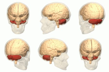
Latin for "little brain". About 10 percent of brain volume and over 50 percent of its neurons.
Its cortex is folded into extremely fine parallel ridges — far finer than the cerebral cortex — and if unfolded it would form a strip about 1 metre long and 5 centimetres wide.
And its circuitry is strikingly uniform. Unlike the cerebral cortex, where different regions look different, the cerebellar cortex has the same microscopic structure everywhere. This has been taken to mean it performs one computation, applied to whatever is fed into it.
What it does
It does not initiate movement. Damage does not cause weakness or paralysis.
It coordinates, times and calibrates movement, comparing the intended movement with the actual movement and correcting the difference.
It receives an enormous input: a copy of every motor command from the cortex, plus position and stretch information from muscles and joints, plus balance information from the inner ear, plus visual information. It compares them and outputs corrections.
This is an error-correction system, and it is why cerebellar damage produces movement that is powerful but wrong — overshooting, wobbling, mistimed.
And it stores learned motor programmes. Riding a bicycle, playing an instrument, a tennis serve — the smooth automatic execution of a practised movement is largely cerebellar. This is why those skills are retained for decades without practice, and why they are lost in cerebellar disease while strength is intact.
Increasingly it is recognised to do the same for cognition. The cerebellum connects extensively with the prefrontal cortex, and damage can produce a "cerebellar cognitive affective syndrome" with impaired planning, flattened affect and language difficulty. The same computation — comparing predicted with actual and correcting — applied to thought rather than movement is the current framing, and it is an active area rather than a settled one.
The signs of cerebellar disease
Memorable as DANISH, and each sign follows from a failure of error correction.
D — Dysdiadochokinesia. Inability to perform rapid alternating movements, such as rapidly turning the hand palm up and palm down. The movements become slow, clumsy and irregular, because each requires stopping one movement and starting its opposite at exactly the right moment.
A — Ataxia. An unsteady, broad-based, staggering gait. Unlike the ataxia of position sense loss, it does not improve with the eyes open — which is what distinguishes it from a dorsal column problem on Romberg's test (Chapter 11.3).
N — Nystagmus. Rhythmic involuntary eye oscillation. Eye movements are movements, and they need the same calibration.
I — Intention tremor. A tremor that appears during a purposeful movement and worsens as the target is approached. Absent at rest.
This is the opposite of the Parkinsonian tremor, which is present at rest and improves with movement — and that single distinction separates the two commonest tremors seen in a clinic.
S — Slurred, scanning speech. Speech broken into separate syllables with uneven emphasis, because speech is a rapid coordinated motor act.
H — Hypotonia and heel–shin test failure, in which the person cannot smoothly run their heel down the opposite shin.
And the past-pointing test — reaching for a target and overshooting, then correcting, then overshooting the other way.
One rule that catches people out: cerebellar signs are on the same side as the lesion, not the opposite side. The cerebellum's connections cross twice, which cancels out. So left cerebellar damage causes left-sided clumsiness — the opposite of the rule for cerebral lesions, and one of the more useful localising facts in neurology.
Causes
Alcohol is the commonest, both acutely and chronically. Acute intoxication produces exactly the DANISH picture — unsteady wide-based gait, slurred speech, nystagmus, and past-pointing — because alcohol depresses cerebellar function preferentially. A roadside sobriety test is a cerebellar examination, and walking heel to toe along a line is a standard cerebellar test used for that reason.
Chronic alcohol use causes permanent degeneration, particularly of the region controlling the legs, which is why the gait is affected out of proportion to the arms.
Stroke in the posterior circulation. Multiple sclerosis, which has a predilection for cerebellar pathways. Tumours — in children, the cerebellum is a common site. Inherited ataxias, including Friedreich's ataxia. Drugs — phenytoin, lithium at toxic levels. Vitamin deficiency — thiamine, giving Wernicke's encephalopathy (Chapter 1.6), and vitamin E.
Posterior fossa problems are dangerous out of proportion to their size, because the space is small and rigid and the brainstem is immediately adjacent. A cerebellar bleed or a swelling cerebellar stroke can compress the brainstem and block cerebrospinal fluid drainage within hours, and it is one of the few strokes where urgent surgery is genuinely life-saving.
The thalamus and hypothalamus
Not brainstem, but sitting immediately above it and worth introducing here.
The thalamus is the relay station. Every sensory pathway except smell synapses here before reaching the cortex.
It is not a passive relay. It gates, filters and modulates — deciding what reaches consciousness and what does not, and it is heavily involved in attention and in arousal.
Thalamic stroke can produce a striking and cruel syndrome: complete sensory loss on one side, followed weeks later by severe spontaneous burning pain in the numb area. Central post-stroke pain is notoriously difficult to treat.
The hypothalamus is the size of an almond and controls, directly or indirectly, an implausible list: body temperature, hunger, thirst, sleep–wake cycles, the autonomic nervous system, the entire endocrine system through the pituitary, and much of emotional and sexual behaviour.
It is the master homeostatic controller (Chapter 4.7), and Chapter 12.2 covers its endocrine role.
Its suprachiasmatic nucleus is the body's master clock, receiving light information directly from the retina and setting the circadian rhythm of every other tissue.
What the next page fixes
Above all of this sits the structure that makes you a person. Chapter 11.5 covers the cerebrum — its lobes, the map of the cortex, what each region does, and what the classic lesions reveal about how thought is organised.