Appearance
7.1 — Blood
You have about 5 litres of blood, roughly 7 to 8 percent of your body weight. It contains around 25 trillion red cells, and you destroy and replace about 2 million of them every second — roughly 200 billion a day, which is why bone marrow is one of the most productive tissues in the body.
Lose 15 percent of your blood volume and your body compensates and you feel light-headed. Lose 30 to 40 percent and you are in shock. Lose more than 40 percent and you will die without transfusion.
What blood is made of
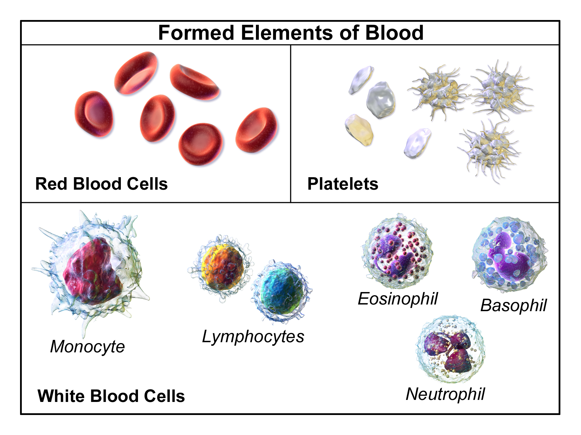
Spin blood in a centrifuge and it separates into layers.
Plasma (about 55 percent) — a straw-coloured fluid, itself about 92 percent water. The remaining 8 percent is proteins, electrolytes, nutrients, hormones and waste products.
The buffy coat (under 1 percent) — a thin pale layer of white cells and platelets.
Red cells (about 45 percent) — the packed layer at the bottom. This percentage is the haematocrit, and it is one of the most useful numbers in a blood test: normally about 40 to 50 percent in men and 36 to 46 percent in women.
Plasma proteins
Around 7 grams per 100 ml, and the three main groups do quite different things.
Albumin (about 60 percent) — made by the liver. Its main jobs are maintaining oncotic pressure and carrying substances that do not dissolve well in water, including many drugs, calcium, bilirubin and fatty acids.
Oncotic pressure deserves explaining properly, because it explains oedema. Albumin cannot leave the capillaries, so it is a solute confined to the blood, and it therefore pulls water in osmotically (Chapter 1.4). Blood pressure pushes fluid out of capillaries; albumin's oncotic pressure pulls it back in. The balance decides how much fluid stays in the tissues.
So a low albumin causes swelling. This is why people with liver disease, who cannot make enough albumin, and people with kidney disease losing albumin in the urine, develop generalised oedema and fluid in the abdomen. And it is why severe protein malnutrition produces the swollen belly of kwashiorkor — a starving child with a distended abdomen is not full, they have no albumin.
Globulins — including the antibodies (Chapter 13.3) and various transport proteins.
Fibrinogen — the soluble precursor of the fibrin mesh in a clot.
Serum is plasma with the clotting factors removed — what is left after blood has clotted. A blood test tube with an anticoagulant gives plasma; one without gives serum. Different tests need different tubes, which is why blood is drawn into several coloured tubes.
Red blood cells
Shape. A biconcave disc, about 7 to 8 micrometres across and 2 micrometres thick, dimpled on both sides.
That shape is engineered for three things. It maximises surface area for a given volume, so gas exchange is fast. It puts every haemoglobin molecule close to the surface, so no oxygen has far to diffuse. And it makes the cell extremely deformable, which matters because capillaries are as narrow as 5 micrometres and the cell must fold to pass.
Structure. A mature human red cell has no nucleus and no mitochondria. It expels them during development.
Both absences are deliberate and both have consequences. No nucleus means more room for haemoglobin — a red cell is about 33 percent haemoglobin by weight — and a more flexible cell. No mitochondria means the cell cannot use any of the oxygen it carries, which would otherwise be a serious loss; it runs entirely on glycolysis (Chapter 1.6).
The cost is that it cannot repair itself or divide. With no nucleus there is no protein synthesis, so as its enzymes wear out it is doomed. Lifespan is about 120 days, after which macrophages in the spleen and liver detect the stiffening membrane and remove it.
Haemoglobin. Four protein chains, each holding a haem group with an iron atom at its centre. Each iron binds one oxygen molecule, so each haemoglobin carries four. A single red cell contains around 270 million haemoglobin molecules, so it carries over a billion oxygen molecules.
Iron is the limiting resource. Total body iron is about 3 to 4 grams, two thirds of it in haemoglobin. The body has no mechanism for excreting excess iron — it only regulates absorption — which is why iron overload from repeated transfusion is so dangerous (Chapter 2.8) and why iron supplements should not be taken without evidence of deficiency.
Breakdown produces the pigments you can see. Haemoglobin is split: the globin is recycled as amino acids, the iron is recovered and reused, and the haem ring becomes bilirubin. Bilirubin is carried to the liver, conjugated, and excreted in bile. This is why bile is green-brown, why stool is brown, and why jaundice — a build-up of bilirubin — turns skin and the whites of the eyes yellow. A bruise changing from purple to green to yellow is the same chemistry happening locally.
Erythropoietin
Red cell production is controlled by a hormone from the kidney. Cells in the kidney sense oxygen delivery, and when it falls they release erythropoietin (EPO), which tells the bone marrow to make more red cells. Production takes about 5 to 7 days.
This is why chronic kidney disease causes anaemia — the kidney fails to produce EPO. It is treated with synthetic EPO, one of the first biotechnology drugs.
It is why living at altitude raises the red cell count, and why altitude training works.
And it is why EPO became one of the most notorious doping agents in endurance sport. Raising the haematocrit increases oxygen delivery — and also increases blood viscosity, which raises the risk of thrombosis. A cluster of deaths among young professional cyclists in the late 1980s and early 1990s is attributed to it.
Anaemia
Anaemia is a reduced capacity to carry oxygen — usually defined by haemoglobin below about 130 g/L in men and 120 g/L in women.
Symptoms are those of reduced oxygen delivery: fatigue, breathlessness on exertion, palpitations, pallor, and headache. Severe cases cause chest pain, because the heart's own supply becomes inadequate.
The single most useful classification is by red cell size (MCV), and it narrows the cause immediately.
Microcytic (small cells) — iron deficiency by far the commonest cause worldwide, then thalassaemia (Chapter 2.8) and chronic disease.
Normocytic (normal size) — acute blood loss, chronic disease, kidney failure, haemolysis, marrow failure.
Macrocytic (large cells) — vitamin B12 or folate deficiency, alcohol, liver disease, hypothyroidism, and some drugs.
Iron deficiency anaemia deserves particular emphasis, because the anaemia is a symptom, not a diagnosis. In a menstruating woman it is usually menstrual loss or pregnancy. In a man, or a postmenopausal woman, iron deficiency anaemia means gastrointestinal blood loss until proven otherwise, and that means colorectal cancer must be excluded. It is one of the standard urgent referral criteria, and treating the anaemia with iron tablets without investigating the cause is a recognised and serious error.
Vitamin B12 deficiency has a specific trap. B12 and folate deficiency both cause macrocytic anaemia, and folate corrects the anaemia in both. But B12 deficiency also causes irreversible neurological damage — degeneration of the spinal cord — and folate does not prevent that. So treating a B12 deficiency with folate makes the blood look better while the nerve damage continues. B12 must always be checked and corrected first.
B12 is absorbed by a specific mechanism: it binds intrinsic factor, made by stomach cells, and the complex is absorbed in the terminal ileum. So it fails in pernicious anaemia (autoimmune destruction of those stomach cells), after stomach surgery, in Crohn's disease affecting the ileum, and after long-term metformin or acid-suppressing drugs. Vegans are at risk because B12 is essentially only in animal products.
Haemolytic anaemias — where cells are destroyed early. Sickle cell (Chapter 2.8), hereditary spherocytosis, G6PD deficiency, autoimmune haemolysis, malaria, and mechanical destruction by a faulty heart valve.
G6PD deficiency is worth knowing because it is drug-related and common. It is X-linked, affects around 400 million people, and is another malaria-protective variant (Chapter 3.3). The enzyme protects red cells against oxidative stress; without it, certain triggers cause sudden haemolysis. The triggers are specific and avoidable: fava beans, some antimalarials including primaquine, sulfonamide antibiotics, nitrofurantoin, and infection. Testing before prescribing primaquine is standard where the deficiency is common.
White blood cells
Around 4,000 to 11,000 per microlitre — about one for every 700 red cells. Part 13 covers what they do; here is what they are and what the counts mean.
Neutrophils (40–70 percent) — the first responders to bacterial infection, short-lived, dying in the process to form pus. A raised neutrophil count suggests bacterial infection.
Lymphocytes (20–45 percent) — B and T cells. A raised lymphocyte count suggests viral infection.
Monocytes (2–10 percent) — become macrophages in tissue.
Eosinophils (1–6 percent) — raised in parasitic infection and in allergy, which is a genuinely useful pairing to remember.
Basophils (under 1 percent) — release histamine.
Neutropenia — a low neutrophil count — is one of the most important results in medicine. Below about 0.5 × 10⁹/L, a person cannot mount a normal response to bacteria. Neutropenic sepsis is a medical emergency: a fever in a patient on chemotherapy must be treated with intravenous antibiotics within an hour, before any results are back, because these patients can deteriorate from mild symptoms to death within hours. The signs of infection are muted precisely because the cells that produce them are absent — there may be no pus, no redness, and few localising signs.
Platelets and clotting
Platelets are cell fragments, 2 to 3 micrometres, budded off from giant marrow cells. Normal count 150,000 to 400,000 per microlitre. Lifespan about 8 to 10 days.
Haemostasis has three stages.
1. Vascular spasm — the damaged vessel constricts immediately, reducing flow.
2. The platelet plug. Platelets stick to exposed collagen at the injury via a bridging protein, become activated, change shape from discs to spiky forms, and release chemicals that recruit and activate more platelets. This is positive feedback (Chapter 4.7), and it produces a plug within seconds.
3. The coagulation cascade — converting that plug into a stable fibrin-reinforced clot.
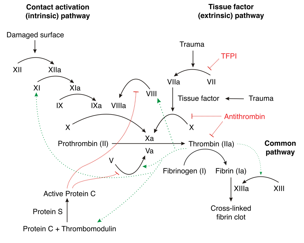
The cascade amplifies. Each activated factor activates many molecules of the next, so a small initiating event produces a clot within seconds.
Vitamin K is required to make factors II, VII, IX and X. This single fact explains several clinical situations at once.
Warfarin works by blocking vitamin K recycling, so those factors are made in a non-functional form. This is why warfarin takes two to three days to work — the existing factors have to be cleared first — and why its effect is reversed by giving vitamin K, which takes hours, or by giving the factors directly, which is immediate.
And it is why warfarin interacts with diet. A sudden increase in green vegetables raises vitamin K intake and reduces the drug's effect. The correct advice is consistency rather than avoidance — eat roughly the same amount of green vegetables, and the dose is adjusted around it.
Newborns are given vitamin K at birth because they have very little: it crosses the placenta poorly, breast milk contains little, and the gut bacteria that produce it have not yet colonised. Without it, a small number of babies develop haemorrhagic disease of the newborn, including intracranial bleeding, and a single injection essentially eliminates it.
Clot breakdown matters too. Plasmin dissolves fibrin, and clot-busting drugs used in stroke and heart attack work by activating this system (Chapter 22.7). The breakdown product D-dimer is measurable, and a normal D-dimer is useful for ruling out a clot — a positive result is far less useful, because it rises in infection, pregnancy, cancer and after surgery.
Two bleeding-disorder categories with different patterns:
Platelet problems cause immediate bleeding from small vessels — bruising, nosebleeds, heavy periods, a pinprick rash (petechiae).
Coagulation factor problems cause delayed, deep bleeding — into joints and muscles, with bleeding restarting hours after an injury (Chapter 2.8).
Disseminated intravascular coagulation (DIC) is the catastrophic version: widespread activation of clotting throughout the circulation, consuming platelets and factors, so that the person clots and bleeds simultaneously. Triggered by sepsis, major trauma, obstetric emergencies and some cancers. Treatment is of the underlying cause; there is no way to treat the DIC itself directly.
What the next page fixes
Blood is useless without something to move it. Chapter 7.2 covers the heart — its position, its four chambers, its valves, what its walls are made of, and why the left ventricle is three times thicker than the right.