Appearance
7.3 — The Conduction System and the ECG
A heart removed from the body keeps beating. A transplanted heart, with every nerve to it cut, beats normally. The heartbeat does not come from the brain — it is generated inside the heart itself, by cells that depolarise spontaneously, and the nervous system only speeds it up or slows it down.
This chapter covers where the beat starts, how it spreads, and how the electrical activity is read off the skin.
Where the beat comes from
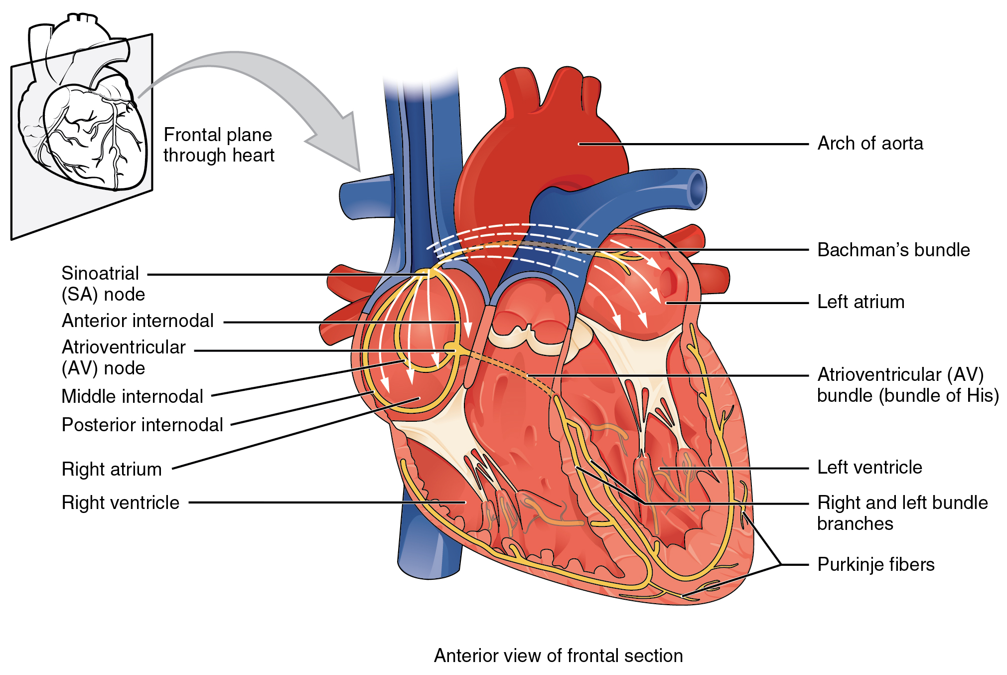
The sinoatrial (SA) node — a patch of specialised cells in the upper right atrium, about 15 millimetres long. These cells have unstable resting membranes: they leak sodium inward continuously, so the membrane drifts upward until it reaches threshold and fires. Then it repolarises and the drift starts again.
That drift is called the pacemaker potential, and it is why the heart beats without instruction. Nothing has to tell it; it simply cannot stay still.
The SA node's intrinsic rate is about 100 beats per minute, and a resting heart rate of 60 to 80 reflects the fact that the vagus nerve is continuously slowing it. This is why cutting the vagus, or blocking it with atropine, raises the heart rate to around 100 — and why a transplanted heart, with no vagal supply, runs at about 90 to 100 at rest.
Several other tissues can also pacemake, at slower intrinsic rates, and this hierarchy is a genuine safety system.
| Site | Intrinsic rate |
|---|---|
| SA node | 60–100 |
| AV node | 40–60 |
| Bundle of His | 40–45 |
| Purkinje fibres | 20–40 |
The fastest pacemaker wins, because it fires before the others have finished drifting. So the SA node normally dominates and the rest are silent backups. If it fails, the AV node takes over at a slower rate — an "escape rhythm" — which is enough to keep a person alive though not well.
The sequence
1. The SA node fires. The impulse spreads across both atria through the muscle itself, cell to cell, taking about 90 milliseconds. Both atria contract.
2. The impulse reaches the atrioventricular (AV) node. The fibrous skeleton (Chapter 7.2) insulates atria from ventricles, so the AV node is the only electrical connection between them.
3. The AV node delays the impulse by about 100 milliseconds, because its cells conduct slowly.
That delay is essential, and it is worth being clear why. It gives the atria time to finish contracting and empty into the ventricles before the ventricles contract. Without it, all four chambers would contract at once, and the atria would be pumping into ventricles that were already squeezing shut.
And the AV node has a second job: it acts as a rate limiter. It cannot conduct faster than about 180 to 200 impulses per minute. This is what protects the ventricles when the atria fire chaotically at 400 to 600 per minute in atrial fibrillation — most impulses are blocked, and only some get through.
4. Down the bundle of His, which pierces the fibrous skeleton, then splits into left and right bundle branches running down either side of the interventricular septum.
5. Into the Purkinje fibres, which spread through the ventricular walls. These conduct at about 4 metres per second, roughly four times faster than ordinary heart muscle, so the whole ventricular mass is activated within about 60 milliseconds.
6. The ventricles contract from the apex upward, which is the right direction — it squeezes blood toward the outlets at the top, exactly as you would squeeze a tube of toothpaste.
The cardiac action potential
Heart muscle's electrical behaviour differs from nerve or skeletal muscle in one crucial way: it has a long plateau.
A nerve action potential lasts about 1 millisecond. A ventricular action potential lasts 200 to 300 milliseconds, because after the initial sodium influx, calcium channels open and calcium enters slowly, holding the membrane depolarised.
Two consequences, and both are protective.
Calcium entry triggers more calcium release from internal stores, which is what couples excitation to contraction in cardiac muscle — and it is why calcium channel blockers reduce the force of cardiac contraction, and why heart muscle depends far more on external calcium than skeletal muscle does.
And the refractory period lasts almost as long as the contraction itself. The cell cannot be re-stimulated until it has nearly finished contracting.
This is why the heart cannot be tetanised. A skeletal muscle stimulated rapidly enough goes into sustained contraction (Chapter 6.1). A heart cannot, and must not — a heart locked in sustained contraction would stop pumping immediately. The long refractory period makes it physically impossible.
The ECG
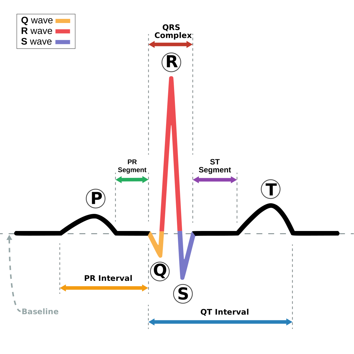
An ECG records the summed electrical activity of the whole heart, detected at the skin. It measures voltage differences between electrodes, and because the body conducts electricity, activity deep in the chest is detectable on the surface.
What each part is:
P wave — atrial depolarisation. Small, because the atria have little muscle.
QRS complex — ventricular depolarisation. Large and sharp, because the ventricles have much more muscle and are depolarised very rapidly by the Purkinje system. Normal duration is under 120 milliseconds — a wide QRS means the impulse is not using the fast conduction pathway.
T wave — ventricular repolarisation.
Atrial repolarisation is not visible — it is buried inside the QRS complex, which is happening at the same time and is much larger.
The intervals, and what each measures:
PR interval (start of P to start of QRS) — normally 120 to 200 ms. This is the AV node delay, so a long PR interval means the AV node is conducting slowly.
QRS duration — under 120 ms. Wide means abnormal ventricular conduction: a bundle branch block, or an impulse originating in the ventricle itself.
QT interval — the total time from ventricular depolarisation to the end of repolarisation. It shortens as heart rate rises, so it is corrected for rate (QTc), normally under about 440 ms in men and 460 ms in women.
A long QT is dangerous and it is one of the most clinically important measurements on the ECG. It means repolarisation is prolonged and uneven, which allows a chaotic ventricular rhythm called torsades de pointes to start — which can degenerate into ventricular fibrillation and cause sudden death.
Long QT can be inherited — several channel gene mutations, and a cause of sudden death in young people, sometimes triggered by exercise or by a sudden loud noise. And it is very commonly caused by drugs: some antiarrhythmics, several antipsychotics, some antibiotics including macrolides and fluoroquinolones, some antidepressants, and some antiemetics. Low potassium and low magnesium make it worse. Combining two QT-prolonging drugs in a patient with low potassium is a recognised route to a cardiac arrest, and it is why these interactions are checked. Chapter 22.13.
The twelve leads
A standard ECG uses ten electrodes to produce twelve views.
Six limb leads view the heart in the vertical (coronal) plane; six chest leads view it in the horizontal plane.
Each lead is a viewpoint, and together they localise where a problem is. This is why an ECG can tell you which coronary artery is blocked before any imaging is done.
| Leads | Region of heart | Artery |
|---|---|---|
| II, III, aVF | Inferior | Right coronary |
| V1–V4 | Anterior/septal | Left anterior descending |
| I, aVL, V5, V6 | Lateral | Circumflex |
In a heart attack, ST elevation in a group of leads identifies the territory, and therefore the artery, and therefore where the interventional cardiologist goes first. Chapter 18.3.
Common abnormalities
Sinus bradycardia — rate under 60 from the SA node. Normal in athletes, whose stroke volume is large enough that a slow rate suffices. Also caused by beta-blockers, hypothyroidism, and raised intracranial pressure.
Sinus tachycardia — over 100 from the SA node. Almost always a response to something rather than a primary problem: fever, pain, anxiety, blood loss, dehydration, hyperthyroidism, sepsis. The correct action is to find the cause, not to slow the heart — slowing a compensatory tachycardia in someone who is bleeding is harmful.
Atrial fibrillation — the atria depolarise chaotically at 400 to 600 per minute instead of contracting in an organised way. On the ECG there is no P wave and the rhythm is irregularly irregular, with no discernible pattern.
Three consequences:
The atria do not contract properly, so the atrial kick — the top-up they give to ventricular filling — is lost. This costs 10 to 30 percent of cardiac output, which matters most in people whose hearts were already struggling.
The ventricular rate is fast and irregular, because whichever impulses get through the AV node do so unpredictably.
And blood stagnates in the non-contracting atrium, particularly in the left atrial appendage, and clots. If the clot leaves, it travels up the aorta and frequently into the brain. Atrial fibrillation causes roughly a fivefold increase in stroke risk and accounts for around 20 to 30 percent of all ischaemic strokes.
Anticoagulation is therefore the central treatment, and the decision is made with a risk score combining age, heart failure, hypertension, diabetes, previous stroke, vascular disease and sex. The strokes caused by AF are on average more severe and more disabling than other strokes, because the clot is larger.
Heart block — impaired conduction through the AV node.
- First degree — PR interval over 200 ms. Every impulse gets through, just slowly. Usually harmless.
- Second degree — some impulses fail to get through. Two patterns, one benign and one not.
- Third degree (complete) — no impulses get through. The atria and ventricles beat completely independently, with the ventricles driven by an escape pacemaker at 20 to 40 per minute. This requires a pacemaker, usually urgently.
Ventricular tachycardia — a fast rhythm originating in the ventricle. Wide QRS, rate 100 to 250. Dangerous, because it fills poorly and can degenerate into fibrillation.
Ventricular fibrillation — completely disorganised ventricular electrical activity. There is no coordinated contraction, so there is no cardiac output. This is cardiac arrest, and it is fatal within minutes.
And it is the rhythm a defibrillator treats. Chapter 23.2 covers the procedure; the mechanism is worth stating here. A defibrillator does not restart the heart. It stops it. A shock depolarises the entire myocardium at once, ending all the chaotic activity, so that the SA node — which is still there and still capable of drifting to threshold — can restart an organised rhythm.
This is why defibrillation only works for shockable rhythms. In asystole — a flat line, no electrical activity — there is nothing to stop, and shocking it does nothing. Television consistently gets this wrong, showing shocks delivered to a flat line. The treatment for asystole is chest compressions and adrenaline.
Pacemakers and defibrillators
A pacemaker delivers small electrical impulses when the heart's own rate is too slow. Leads are passed through a vein into the heart and the generator sits under the skin below the collarbone. Modern devices sense the heart's own activity and only fire when needed, and can adjust rate with activity.
An implantable cardioverter defibrillator (ICD) is a pacemaker that can also deliver a shock if it detects ventricular fibrillation or fast ventricular tachycardia. Fitted to people at high risk of sudden cardiac death — after a cardiac arrest, in severe heart failure, or in inherited arrhythmia syndromes.
A magnet placed over a pacemaker or ICD changes its behaviour, which is used in emergencies — for instance to stop an ICD delivering repeated inappropriate shocks to a conscious patient, which is extremely distressing.
And an ICD must be deactivated at the end of life, or a dying patient receives repeated painful shocks. This is a genuine and frequently overlooked part of palliative care planning.
What the next page fixes
The electrical events cause mechanical ones. Chapter 7.4 covers the cardiac cycle — what the pressures and volumes are doing at every moment, how the heart automatically matches its output to what returns to it, and what determines how much blood is pumped per minute.