Appearance
5.1 — Bone as Living Tissue
A museum skeleton is dry, white and inert, and it gives entirely the wrong impression. Living bone is about 10 percent water, richly supplied with blood, packed with nerves, and completely rebuilt roughly every ten years. It is one of the most metabolically active tissues in your body, it manufactures every blood cell you have, and it is the bank account from which your bloodstream withdraws calcium whenever your diet falls short.
You have 206 bones as an adult and around 270 at birth — the difference is fusion, mostly in the skull and pelvis and sacrum.
What bone is made of
Bone is a composite material, and its properties come from combining two things that fail in opposite ways.
About 65 percent by weight is mineral — mostly hydroxyapatite, a crystal of calcium and phosphate with the formula \mathrm{Ca_{10}(PO_4)_6(OH)_2}. Mineral is hard and resists compression, and on its own it is brittle: it shatters rather than bends, like chalk.
About 25 percent is organic matrix, almost all type I collagen (Chapter 4.2). Collagen is flexible and resists pulling, and on its own it is soft.
About 10 percent is water.
Together they give something neither could be alone. This is exactly the principle of reinforced concrete: concrete is strong in compression and weak in tension, steel rods are strong in tension, and the composite is strong in both. Bone is the biological version, and it beats concrete comfortably — bone's compressive strength is around 170 megapascals, comparable to some grades of cast iron, at a fraction of the density.
You can demonstrate the two components separately, and the experiment is memorable. Soak a bone in acid and the mineral dissolves; what remains is collagen, and the bone becomes rubbery enough to tie in a knot. Bake a bone and the collagen burns away; what remains is mineral, and it crumbles at a touch. Diseases that mimic each of these exist. Rickets and osteomalacia are failures of mineralisation, giving soft bones that bend. Osteogenesis imperfecta is a collagen defect, giving bones that snap (Chapter 4.2).
Compact and spongy bone

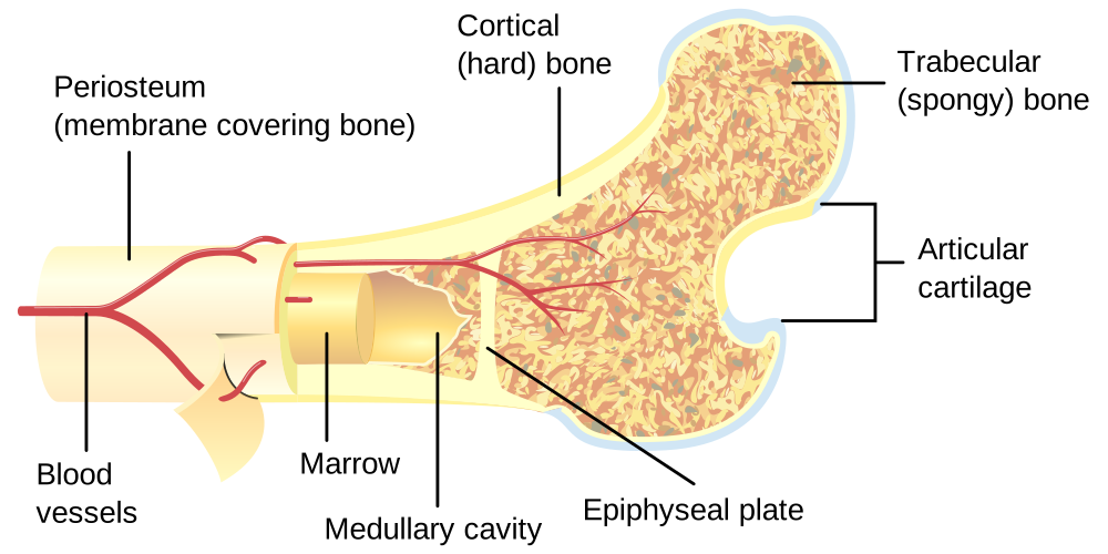
Compact (cortical) bone is dense, forms the outer shell of every bone and the shaft of long bones, and makes up about 80 percent of skeletal mass. It carries the load.
Spongy (trabecular or cancellous) bone is an open lattice of struts found at the ends of long bones and inside flat bones. It is only 20 percent of mass but has a far greater surface area, which matters for two reasons: it is where most of the mineral exchange with the blood happens, and it is where the marrow lives.
The lattice is engineered. The struts align with the lines of principal stress through the bone — an arrangement worked out in the nineteenth century when an anatomist showed the trabecular pattern in the top of the femur to an engineer, who recognised it as the stress pattern in a loaded crane arm. This is why spongy bone gives strength at a fraction of the weight, and why a femur can be hollow.
The microscopic structure
Compact bone is built from repeating cylinders called osteons, each about 200 micrometres across, running along the bone's long axis.
At the centre of each osteon runs a central (Haversian) canal carrying a blood vessel and a nerve. Around it, matrix is laid down in concentric rings, and the collagen fibres in adjacent rings run in different directions — the same trick used in plywood and in vehicle tyres, and for the same reason: it resists twisting and stops cracks running straight through.
Bone cells sit trapped in small spaces between the rings, each connected to its neighbours by hair-thin channels through which fine cell processes reach. No bone cell is more than about 0.2 millimetres from a blood vessel, because nutrients cannot diffuse through mineralised matrix — they have to travel through those channels.
The four cell types
Osteoblasts — the builders. They secrete collagen and the other matrix proteins, then control mineralisation. Derived from the same stem cells as fat and cartilage cells.
Osteocytes — the sensors. An osteoblast that becomes trapped in the matrix it has just laid down becomes an osteocyte. They are 90 to 95 percent of all bone cells and they live for decades. For a long time they were assumed to be retired osteoblasts doing nothing. They are the opposite: they are the strain sensors that tell the whole system where bone is needed. Their long processes detect the movement of fluid through the channels when the bone bends, and they signal accordingly.
Osteoclasts — the demolishers. Large multinucleated cells formed by fusion, derived from the same lineage as white blood cells. An osteoclast seals itself to the bone surface and pumps acid into the sealed space, dissolving the mineral, then releases enzymes that digest the collagen. It is essentially a cell that creates a private stomach against the bone surface.
Lining cells cover resting bone surfaces and control what reaches them.
Remodelling: why bone is never finished
About 10 percent of your skeleton is replaced every year. Trabecular bone turns over faster than cortical — roughly every 3 to 4 years against 10 or more. Over a lifetime you replace the whole thing several times.
The cycle takes 3 to 6 months in one location:
- Osteocytes detect microdamage or a change in strain and signal.
- Osteoclasts arrive and resorb bone, taking about 2 to 3 weeks.
- Osteoblasts arrive and lay down new matrix, taking 3 to 4 months.
- The matrix mineralises over months more.
Note the asymmetry: destruction is fast, construction is slow. That is why anything increasing the number of remodelling sites — which is what happens after menopause — causes net bone loss, and why a drug that reduces remodelling can increase bone density simply by letting existing sites finish filling in.
Why remodel at all? Three reasons.
To repair microdamage. Ordinary loading produces microscopic cracks continuously. Left alone they would accumulate and the bone would eventually fail — which is exactly what a stress fracture is: damage arriving faster than remodelling can repair it.
To adapt to load. Wolff's law, stated in 1892: bone adapts to the forces placed on it. Load a bone more and it thickens; unload it and it thins.
The evidence is dramatic. The playing arm of a professional tennis player has 20 to 30 percent more cortical bone than the other arm — the same person, the same genes, the same diet, different load. Astronauts lose 1 to 2 percent of bone mass per month in weightlessness, concentrated in the weight-bearing bones of the legs and spine, and it does not fully recover on return. Prolonged bed rest does the same.
And this is why exercise is the only intervention that builds bone rather than merely preserving it. The stimulus is strain, so the effective exercises are impact and resistance — running, jumping, weight training. Swimming and cycling are excellent for the heart and do very little for bone, because the skeleton is unloaded. Chapter 24.6.
To manage calcium. This is the third job and it overrides the other two.
Bone as the calcium bank
Your blood calcium is held between 2.2 and 2.6 mmol/L, and the tolerance is tight because calcium controls nerve firing, muscle contraction, clotting and hormone release. Too low and nerves fire spontaneously, causing muscle spasms and, at the extreme, laryngeal spasm and death. Too high and nerves become sluggish, causing confusion, constipation, kidney stones and cardiac arrhythmia.
About 99 percent of your body's calcium — around a kilogram — is in your bones, and the skeleton is treated as a reserve.
Parathyroid hormone (PTH) is released when blood calcium falls, and it raises it three ways: it stimulates osteoclasts to dissolve bone, it makes the kidney retain calcium, and it activates vitamin D, which increases absorption from the gut.
Calcitonin, from the thyroid, does the reverse, though its role in adult humans is minor.
Vitamin D is required for calcium absorption, and it is not really a vitamin — it is a hormone your skin makes from cholesterol when ultraviolet light hits it. It is then activated in two steps, in the liver and then the kidney.
This has consequences you can act on. Deficiency is extremely common at high latitudes, in people with darker skin living far from the equator, in those who cover their skin, and in the housebound. Severe deficiency causes rickets in children — soft bones bending under load, giving the bowed legs and deformed ribcage that were endemic in industrial cities before fortification — and osteomalacia in adults, causing bone pain and muscle weakness. And because the final activation step happens in the kidney, chronic kidney disease causes a bone disease as a direct consequence (Chapter 21.3).
The system's priority order is worth naming plainly: blood calcium wins over bone strength, every time. A body facing chronic calcium shortage will dismantle the skeleton to keep the blood level correct, because a nerve failing today kills faster than a fracture next year.
Marrow: the blood factory
The spaces inside spongy bone contain marrow, and it comes in two forms.
Red marrow makes blood — around 200 billion red cells, 10 billion white cells and 400 billion platelets every day. In a child it fills nearly every bone. In an adult it is confined to the pelvis, sternum, ribs, vertebrae, skull and the upper ends of the femur and humerus, and the rest has been replaced by fat.
That distribution is exactly why a bone marrow biopsy is taken from the back of the pelvis, and why a sternal or pelvic sample is what a haematologist asks for.
Yellow marrow is fat, and it can convert back to red marrow when demand is high — after severe blood loss or in chronic anaemia.
And in an emergency where no vein can be found, fluid and drugs can be given directly into the marrow cavity — intraosseous access, usually into the upper tibia. The marrow drains into the venous system, so the drug reaches the circulation within seconds, and it works at essentially the same speed as an intravenous line. It is now standard practice in paediatric resuscitation and in trauma, and it exists because bone is not a solid rod (Chapter 23.1).
How bones form and grow
Two routes, and the difference shows up in fracture healing.
Intramembranous ossification — bone forms directly from connective tissue with no cartilage stage. The flat bones of the skull, the face and the clavicle form this way.
Endochondral ossification — a cartilage model is laid down first and then replaced by bone. Every long bone forms this way, and the process explains growth.
Growth in length happens at the growth plate — a disc of cartilage between the shaft and the end of a long bone. Cartilage cells multiply on the side facing the end, enlarge, and are replaced by bone on the side facing the shaft. So the bone lengthens by pushing its end away, while the plate itself stays the same thickness.
Growth stops when the plate closes — the cartilage is entirely replaced by bone and no further lengthening is possible. Oestrogen closes growth plates, in both sexes, and this explains several things at once. It is why girls stop growing earlier than boys, because oestrogen rises earlier in puberty. It is why the growth spurt precedes the stop. And it is why a rare condition in which a man cannot respond to oestrogen results in someone who simply keeps growing into adulthood.
Growth plates are the weak point in a child's skeleton. A force that would sprain an adult's ligament instead fractures through the growth plate in a child, because the plate is weaker than the ligament. A growth plate fracture can arrest growth on one side and cause progressive deformity, so these injuries are classified carefully and followed up, and "it's just a sprain" is a diagnosis to be suspicious of in a child.
Growth in width happens at the surface, with osteoblasts adding bone on the outside while osteoclasts widen the marrow cavity from within — so the bone gets wider without getting proportionally heavier.
And bone age is readable. The sequence in which growth centres appear and fuse is consistent enough that an X-ray of a child's hand gives a skeletal age, which is compared with chronological age to investigate growth disorders — and, more controversially, to estimate the age of people with no documentation.
What bones tell a forensic scientist
Bone survives when nothing else does, and it records a great deal.
Sex, from the pelvis primarily (Chapter 5.6), and from skull features, with around 95 percent accuracy from a complete pelvis.
Age, from growth plate fusion in the young, and from degeneration of the pubic joint surface and the rib ends in adults, with accuracy that falls as age increases.
Height, from long bone lengths using regression formulae — the femur gives the best estimate.
Old injuries. A healed fracture is visible for life.
Disease. Syphilis, tuberculosis, rickets, scurvy, arthritis and many cancers leave characteristic marks.
And diet and origin, from chemistry. Strontium and oxygen isotopes in tooth enamel reflect the water and geology where a person grew up, because enamel is laid down in childhood and never remodelled. Bone collagen, which does turn over, reflects the last decade or so. So teeth say where you were born and bones say where you have been living recently — which has been used both in archaeology and in identifying unknown remains.
What the next page fixes
That is bone as a material and as an organ. From here the Part goes region by region, and Chapter 5.2 starts at the top: the skull, which is 22 bones doing three quite different jobs — protecting the brain, holding the sense organs, and chewing — and which is where several of the most instructive fractures in medicine occur.